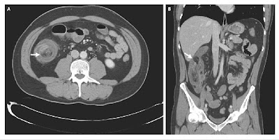
Ileocecal Intussusception
A 45-year-old man with no notable medical or surgical history presented with a 24-hour history of intense pain in the right side of the abdomen with associated nausea and vomiting. He reported having had similar but much less severe episodes during the previous 6 months. Results of initial laboratory tests were unrevealing.
Physical examination showed moderate abdominal distention. Computed tomographic scans of his abdomen revealed an ileocecal intussusception (Panel A, arrow) with a pathologic mass, 2.5 cm in diameter, at the apex, also known as the lead point (Panel B, arrow). Diagnostic laparoscopy was performed, and the diagnosis of intussusception was confirmed.
Laparoscopically assisted ileocecal resection with primary anastomosis was performed. Gross inspection of the specimen showed a pedunculated lipoma within the terminal ileum. The patient had a rapid recovery, with complete resolution of his symptoms.
Labels: CASES, Emergency Medicine, GIT SURGERY, RADIOLOGY


<< Home