Thursday, January 6, 2011
Wednesday, January 5, 2011
Indications for OPG requests
An OPG or "ORTHOPANTOGRAM", gives a panoramic view of the mandible and teeth.
It is performed using a technique called "tomography". The X-ray tube moves around the head, the x-ray film moves in the opposite direction behind your head. This generates an image slice where the mandible and teeth are in focus, and the other structures are blurred.
Dental Disease
* Caries - appear as different shaped areas of ...........Sunday, January 2, 2011
Pneumocephalus
X-ray showing nasal and orbital fractures and pneumocephalus
Pneumocephalus is the presence of air in the cranial vault. It is usually associated with neurosurgery, barotrauma, basilar skull fractures, sinus fractures, nasopharyngeal tumor invasion and meningitis..........Labels: Emergency Medicine, RADIOLOGY
Supraspinatus Pathology by Ultrasound
This video details the complex structures of the human shoulder and how proper and careful ultrasound scanning techniques can identify tendon and rotator cuff tears, as well as avoid false positive and false negative tear diagnoses.
Labels: RADIOLOGY
Saturday, January 1, 2011
Contraindications to MRI procedure
The principal contraindications of the MRI procedure are mostly related to the presence of metallic implants in a patient. The risks of MRI scans increase with the used field strength. In general, implants are becoming increasingly MR safe and an individual evaluation is carried out for each case.
# Intra-orbital metal fragments
# Any electrically, magnetically or mechanically activated implants (including cardiac pacemakers, biostimulators, neurostimulators, cochlear implants, and hearing aids).
-other pacemakers, e.g. for the carotid sinus
-insulin pumps and nerve stimulators
-lead wires or similar wires (MRI Safety risk)
-prosthetic heart valves (in high fields, if dehiscence is suspected)
-haemostatic clips (body)
-non-ferromagnetic stapedial implants
Absolute Contraindications for the MRI scan:
# Intracranial aneurysm clips (Unless the referring physician is certain that it is made of nonferromagnetic material such as titanium).# Intra-orbital metal fragments
# Any electrically, magnetically or mechanically activated implants (including cardiac pacemakers, biostimulators, neurostimulators, cochlear implants, and hearing aids).
Relative Contraindications for the MRI scan:
-cochlear implants-other pacemakers, e.g. for the carotid sinus
-insulin pumps and nerve stimulators
-lead wires or similar wires (MRI Safety risk)
-prosthetic heart valves (in high fields, if dehiscence is suspected)
-haemostatic clips (body)
-non-ferromagnetic stapedial implants
Labels: RADIOLOGY
Thursday, December 30, 2010
Differences between sutures and fractures in skull x-ray
Linear fracture results from low-energy blunt trauma over a wide surface area of the skull. It runs through the entire thickness of the bone and, by itself, is of little significance except when it runs through a vascular channel, venous sinus groove, or a suture. In these situations, it may cause epidural hematoma, venous sinus thrombosis and occlusion, and sutural diastasis, respectively. Differences between sutures and fractures are summarized in this Table
Click here for enlargment
Labels: RADIOLOGY
Sunday, December 26, 2010
Nutcracker phenomenon
It results most commonly from the compression of the left renal vein between the abdominal aorta (AA) and superior mesenteric artery (SMA), although other variants exist. The name derives from the fact that, in the sagittal view, the SMA and AA (with some imagination) appear to be a nutcracker crushing a nut (the renal vein).
There is a wide spectrum of clinical presentations and diagnostic criteria are not well defined resulting in frequently delayed or incorrect diagnosis. This condition is not to be confused with superior mesenteric artery syndrome, which is the compression of the third portion of the duodenum by the SMA and the AA.
This Computed tomography for a 52-year-old woman with mild episodic gross hematuria revealed compression of the left renal vein between the superior mesenteric artery (SMA) and the aorta (Ao) before the vein merges into the inferior vena cava (IVC). This compression caused marked dilatation of the distal part of the renal vein (RV). The renal venous congestion caused hematuria, presumably through the rupture of submucosal veins into the renal pelvis. No treatment was given. Entrapment of the renal vein is a known cause of hematuria.
There is a wide spectrum of clinical presentations and diagnostic criteria are not well defined resulting in frequently delayed or incorrect diagnosis. This condition is not to be confused with superior mesenteric artery syndrome, which is the compression of the third portion of the duodenum by the SMA and the AA.
This Computed tomography for a 52-year-old woman with mild episodic gross hematuria revealed compression of the left renal vein between the superior mesenteric artery (SMA) and the aorta (Ao) before the vein merges into the inferior vena cava (IVC). This compression caused marked dilatation of the distal part of the renal vein (RV). The renal venous congestion caused hematuria, presumably through the rupture of submucosal veins into the renal pelvis. No treatment was given. Entrapment of the renal vein is a known cause of hematuria.
Labels: NEPHROLOGY, RADIOLOGY
Saturday, December 25, 2010
Testicular microliths in a new born
The above ulltrasound images and Color Doppler image show testicular microliths (left testis) in a new born baby.
Testicular calcifications can occur in any age group; however the occurrence of this condition in a newborn is very rare. The color Doppler study showed normal vascularity in the affected testis.
Such cases need to be followed up for changes such as germ cell tumor and more benign conditions such as epididymitis, orchitis and varicocele as well as testicular atrophy. Studies show possible link between Testicular microlithiasis in infants and Down's syndrome.
Labels: PEDIATRiCS, RADIOLOGY
Thursday, December 23, 2010
Osteoporosis of aging (senile or postmenopausal osteoporosis)
Most common form of generalized osteoporosis. As a person ages, the bones lose density and become more brittle, fracturing more easily and healing more slowly. Many elderly persons are also less active and have poor diets that are deficient in protein. Females are affected more often and more severely than males, as postmenopausal women have deficient gonadal hormone levels and decreased osteoblastic activity.
Osteoporosis of aging. Generalized demineralization of the spine in a postmenopausal woman. The cortex appears as a thin line that is relatively dense and prominent (picture-frame pattern).
Labels: ENDOCRINE, ORTHOPEDICS, RADIOLOGY
Anatomy on Thyroid sonography
The anterior neck is depicted rather well with standard gray scale sonography. (FIGURE 1) The thyroid gland is slightly more echo-dense than the adjacent structures because of its iodine content. It has a homogenous ground glass appearance. Each lobe has a smooth globular-shaped contour and is no more than 3 - 4 centimeters in height, 1 - 1.5 cm in width, and 1 centimeter in depth. The isthmus is identified, anterior to the trachea as a uniform structure that is approximately 0.5 cm in height and 2 - 3 mm in depth.
The pyramidal lobe is not seen unless it is significantly enlarged. In the female, the upper pole of each thyroid lobe may be seen at the level of the thyroid cartilage, lower in the male. The surrounding muscles are of lower echogenicity than the thyroid and tissue planes between muscles are usually identifiable. The air-filled trachea does not transmit the ultrasound and only the anterior portion of the cartilaginous ring is represented by dense, bright echoes. The carotid artery and other blood vessels are echo-free unless they are calcified. The jugular vein is usually in a collapsed condition and it distends with a Valsalva maneuver. There are frequently 1-2 mm echo-free zones on the surface and within the thyroid gland that represent blood vessels. The vascular nature of all of these echoless areas can be demonstrated by color Doppler imaging to differentiate them from cystic structures (10-11).
Lymph nodes may be observed and nerves are generally not seen. The parathyroid glands are observed only when they are enlarged and are less dense ultrasonically than thyroid tissue because of the absence of iodine. The esophagus may be demonstrated behind the medial part of the left thyroid lobe, especially if it is distended by a sip of water. (FIGURE 2)
The pyramidal lobe is not seen unless it is significantly enlarged. In the female, the upper pole of each thyroid lobe may be seen at the level of the thyroid cartilage, lower in the male. The surrounding muscles are of lower echogenicity than the thyroid and tissue planes between muscles are usually identifiable. The air-filled trachea does not transmit the ultrasound and only the anterior portion of the cartilaginous ring is represented by dense, bright echoes. The carotid artery and other blood vessels are echo-free unless they are calcified. The jugular vein is usually in a collapsed condition and it distends with a Valsalva maneuver. There are frequently 1-2 mm echo-free zones on the surface and within the thyroid gland that represent blood vessels. The vascular nature of all of these echoless areas can be demonstrated by color Doppler imaging to differentiate them from cystic structures (10-11).
Lymph nodes may be observed and nerves are generally not seen. The parathyroid glands are observed only when they are enlarged and are less dense ultrasonically than thyroid tissue because of the absence of iodine. The esophagus may be demonstrated behind the medial part of the left thyroid lobe, especially if it is distended by a sip of water. (FIGURE 2)
Figure 1. Sonogram of the neck in the transverse plane showing a normal right thyroid lobe and isthmus. L=small thyroid lobe in a patient who is taking suppressive amounts of L-thyroxine, I=isthmus, T=tracheal ring ( dense white arc is calcification, distal to it is artefact), C=carotid artery ( note the enhanced echoes deep to the fluid-filled blood vessel), J=jugular vein, S=Sternocleidomastoid muscle, m=strap muscle.
Figure 2. Sonogram of the left lobe of the thyroid gland in the transverse plane showing a rounded lobe of a goiter. L=enlarged lobe, I= widened isthmus, T=trachea, C=carotid artery ( note the enhanced echoes deep to the fluid-filled blood vessel), J=jugular vein, S=Sternocleidomastoid muscle, m=strap muscles, E=esophagus.
Wednesday, December 22, 2010
Complementary ultrasound to mammography ; Why?
Breast cancer can lie hidden from mammography in women with dense breast tissue, and thus the role of ultrasound is implemented to evaluate the texture and anatomical structures within the breast. Women most likely to have dense breasts are younger, premenopausal women and postmenopausal women who are taking hormone replacement therapy.
Breast ultrasound is used as a complementary examination to mammography in the following situations:
* Evaluation of dense breast tissue.
* Evaluation of a mass demonstrated on mammography.
* Guidance of biopsy needle or needle localization (see cyst aspiration image)
Breast ultrasound is used as a complementary examination to mammography in the following situations:
* Evaluation of dense breast tissue.
* Evaluation of a mass demonstrated on mammography.
* Guidance of biopsy needle or needle localization (see cyst aspiration image)
Labels: GENERAL SURGERY, RADIOLOGY
Tuesday, December 21, 2010
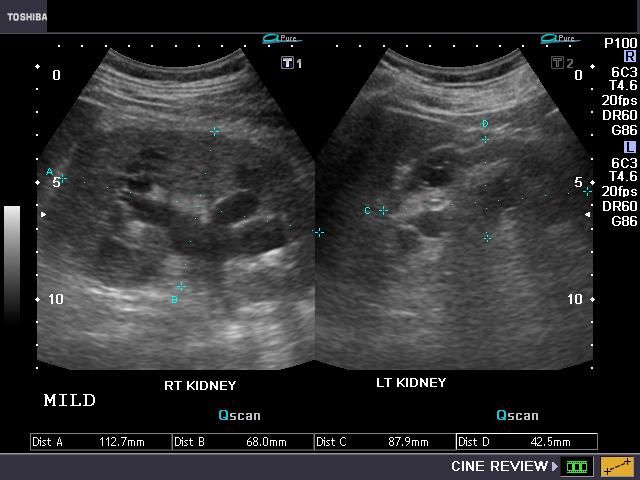
Grades of hydronephrosis (on Ultrasound imaging):
Monday, December 20, 2010
A case of Giant Left Atrium
An 83-year-old woman with long-standing atrial fibrillation who had previously undergone atrioventricular nodal ablation and pacemaker placement presented with symptoms of progressive heart failure.
Physical examination was notable for elevated jugular venous pressure, precordial lift, a grade 2/6 holosystolic murmur at the sternal border and apex, hepatomegaly, ascites, and severe lower-extremity edema.
Laboratory evaluation revealed a creatinine level of 1.4 mg per deciliter (124 µmol per liter), an albumin level of 3.6 g per deciliter, and a brain natriuretic peptide level of 526 pg per milliliter (normal range, 0 to 100 pg per milliliter); liver function was normal.
Chest radiography
An echocardiogram
An esophagogram
The patient was discharged home on medical management after prolonged diuresis.
Labels: CARDIOLOGY, CASES, RADIOLOGY
Friday, December 17, 2010
Patient Preparation and Position for the Ultrasound Examination
Patient Preparation.
The ultrasound examination is most effective if the patient has been NPO for at least 6 hours. This allows the biliary system to be distended and easily imaged by the sonographer. When a patient is fasting there is a a decreased opportunity for gas to accumulate within the colon; gas prohibits the passage of the sound and thus limits visualization of abdominal structures. If the patient is able to consume liquids and the pancreas is not well-visualized, the administration of 32 oz. of water may be given to fill the stomach and duodenum to better delineate the pancreas.The kidneys are best imaged when the patient is fully hydrated, therefore no patient preparation is necessary when only the kidneys are examined. Full hydration will also enable the sonographer to image the distended urinary bladder.
Patient Position.
The position of the patient for the general abdominal scan is usually supine for the initial images. The patient is then rolled into various degrees of obliquity to better demonstrate the biliary system, pancreas, liver, kidneys, or spleen. If the scanning plane is oblique, the sonographer should indicate the change of position on the documented image without specifying the exact degree of obliquity. The same would apply if the patient were in a lateral, upright, or prone position.Labels: RADIOLOGY
Thursday, December 16, 2010
Color Atlas of Ultrasound Anatomy
Publisher: Thieme | ISBN: 1588902811 | edition 200| PDF | 296 pages | 20,9 mb
This brilliant pocket guide helps you to grasp the connection between three-dimensional organ systems and their two-dimensional representation in ultrasound imaging. Through dynamic illustrations and clarifying text, it allows you to: - Recognize, name, and confidently locate all organs, landmarks, and anatomical details of the abdomen -Examine all standard planes, including transverse and longitudinal scans for regions of sonographic interest (including the thyroid gland) - Understand topographic relationships of organs and structures in all three spatial planes This invaluable text is ideal for the beginner, providing a rapid orientation to all key topics.
Labels: FREE MEDICAL BOOKS, RADIOLOGY
Sunday, December 12, 2010
Friday, December 10, 2010
Simple identification of Giant cell tumor in X-ray
Lytic lesion in the end of a long bone of a young adult after epiphyseal closure. Usually asymptomatic, but may be associated with intermittent dull pain and a palpable tender mass and predispose to pathologic fracture. Approximately 20% are malignant (best seen as tumor extension through the cortex and an associated soft-tissue mass on CT). There is much overlap in the radiographic appearance of benign and malignant lesions.
Imaging Findings :
Eccentric lucent metaphyseal lesion that may extend to the immediate subarticular cortex of a bone but does not involve the joint. Expansion toward the shaft produces a well-demarcated lucency, often with cortical expansion but without a sclerotic shell or border. Typically involves the distal femur, proximal tibia, distal radius, or ulna.Giant cell tumor of the distal femur. Typical eccentric lucent lesion in the metaphysis extends to the immediate subarticular cortex. The surrounding cortex, though thinned, remains intact.
Malignant giant cell tumor. The tumor has caused cortical disruption, extends outside the host bone, and has an illdefined margin.
Labels: ORTHOPEDICS, RADIOLOGY
Thursday, December 9, 2010
Pelvic Spleen

A healthy, 23-year-old, nulliparous woman presented with lower abdominal pain, which she reported having had intermittently for the previous year. She had no other symptoms of gastrointestinal distress. A peripheral-blood smear showed no Howell–Jolly bodies.
Physical examination revealed a palpable mass in the suprapubic region. Abdominal ultrasonography revealed displacement of the spleen from its normal position and a homogeneous soft-tissue mass (measuring 11.0 by 9.3 by 4.2 cm) in the pelvis. Imaging of the liver and spleen after injection of technetium-99m sulfur colloid (Panel A) revealed a normal liver and a well-defined area showing abnormal accumulation of radiotracer (arrow) in the lower abdomen.
Multislice computed tomography, with three-dimensional reconstruction of a coronal image (Panel B), revealed the position of the spleen in the pelvis (black arrow), with torsion of the elongated pedicle (white arrow).
Wandering spleen, or pelvic spleen, is an uncommon condition associated with laxity or malformation of the suspensory ligaments of the spleen. Splenic torsion and infarction are potential complications. The principal therapeutic options are splenopexy and splenectomy. After discussion of treatment options, the patient declined surgical intervention and has continued to do so in follow-up over the past 3 years.
Labels: CASES, GIT SURGERY, HEAMATOLOGY, RADIOLOGY