The CliffsTestPrep series offers full-length practice exams that simulate the real tests; proven test-taking strategies to increase your chances at doing well; and thorough review exercises to help fill in any knowledge gaps.
CliffsTestPrep NCLEX-RN is a complete study guide to help you prepare for — and pass — the new NCLEX-RN (the National Council Licensure Examination that is required to obtain a license as a registered nurse). This book contains eight chapters; each chapter contains questions based on the newest version of the exam. Inside this test prep tool, you’ll find
* Three practice exams with answers and explanations
* Coverage of exam areas in terms of what to expect, what you should know, what to look for, and how you should approach each part
* Guidance on how to focus your review of specific subjects to make the most of your study time
* Introduction to the format and scoring of the exam, overall strategies for answering multiple-choice questions, and questions commonly asked about the NCLEX
This book will help you understand the types of questions that will test your knowledge of several basic areas, such as basic patient care and comfort (hygiene, nutrition, mobility/immobility, and more). In addition, you’ll prepare to show what you know about
* Chemical dependency
* Safety and infection control
* Pharmacological dosage calculations
* Diagnostic and laboratory tests
* Infectious diseases and medical emergencies
With guidance from the CliffsTestPrep series, you’ll feel at home in any standardized-test environment!
Your complete guide to a higher score on the NCLEX-RN
Why CliffsTestPrep Guides?
Go with the name you know and trust
Get the information you need—fast!
Written by test prep specialists
About the contents:
Introduction
* How CAT (Computerized Adaptive Testing) exams work
* Overview of the test
* Successful test-taking strategies
* Effective study tips and techniques
Part I: Practice Questions in 8 Subject Areas
* More than 3,200 questions with answers and detailed explanations
* Client Needs categories: Safe and Effective Care Environment, Health Promotion and Maintenance, Psychosocial Integrity, and Physiological Integrity
* Six subcategories
* Concepts integrated throughout: Nursing Process, Caring, Communication and Documentation, Teaching and Learning
Part II: 5 Full-Length Practice Exams with Answers and Detailed Explanations
* Answers facilitate review
* Detailed explanations demonstrate the reasoning processCD-ROM with All Questions in the Book, Including 5 Practice Tests, Answers, and Detailed Explanations
* Test mode allows immediate feedback on a question-by-question basis
* Provides experience answering questions electronically
Test Prep Essentials from the Experts at CliffsNotes®
An American BookWorks Corporation Project
HERE - OR -
HERELabels: FREE MEDICAL BOOKS, Nursing
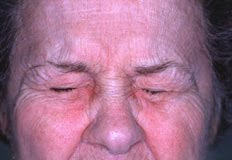
 Blepharospasm is an abnormal tic, spasm, or twitch of the eyelid. It is sometimes referred to as Benign Essential Blepharospasm. Focal dystonia is another phrase for this condition, which involves a involuntary muscle contraction around the eyes. The cause can be fatigue, irritant or caffeine. The symptoms can last for a few days, but commonly disappear without treatment. Severe cases can be chronic.


