all you need to enjoy medicine. Daily medical Videos,Cases,Diagrams,
Animation,Articles,Free medical books and news.
Friday, January 14, 2011
Basosquamous Carcinoma in the Temple
This photo shows large, crusted tumor on the temple that has clinical features of both squamous cell carcinoma (scaly crust), and basal cell carcinoma (translucency in some areas)........
Wolfflin spots are the whitish clumps of connective fibers found in some irides (mostly blue) near the periphery. These are somewhat distinct from Brushfield spots.............
Traumatic exophthalmos develops as a retrobulbar hematoma that pushes the eyeball globe outward. Patient presents with periorbital edema, ecchymosis, a marked decrease in visual acuity, and an afferent pupillary defect in the involved eye. Sometimes the exophthalmos may be obscured by ..........
Port wine stains (PWS) are almost always evident at birth and are a capillary malformation. They are usually sporadic, although some occur in families in an apparently autosomal dominant inheritance. They are initially pink or red, and grow in proportion to infant growth. Although they may appear to lighten in the first few months, they generally darken after this.
They can be associated with other skin anomalies such as extensive mongolian spots. An important association is Sturge-Weber syndrome ( PWS+mental retardation ): PWS involving the forehead (V1 area of the trigeminal nerve), eye abnormalities (choroidal vascular abnormalities, glaucoma), and leptomeningeal and brain abnormalities (vascular malformations, calcification, or cerebral atrophy). Another association is Klippel-Trenaunay syndrome, with a capillary malformation and overgrowth of the soft tissue and bone of the affected limb.
Treatment of PWS is possible using a laser, although improvement rather than complete resolution is most common.
After years of gouty attacks, patients will develop a chronic arthritis resulting in bone and cartilage destruction and deformity. Uric acid crystals deposit within and surrounding the joint causing a chronic destructive inflammatory process. X-rays characteristically show well demarcated "punched out" erosions.
Tophi at helix of ear.
Tophi or deposits of uric acid crystals are often found around joints, in the olecranon bursa or at the pinna of the ear.
X-ray showing soft tissue swelling and erosion of 2nd PIP.
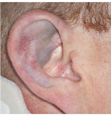
Battle's sign, also called mastoid ecchymosis : consists of bruising over the mastoid process (just behind the auricle), as a result of extravasation of blood along the path of the posterior auricular artery.
It is an indication of fracture of the base of the posterior portion of the skull, and may suggest underlying brain trauma.
An omphalocele is a birth defect in which the infant's intestine or other abdominal organs stick out of the belly button (navel). In babies with an omphalocele, the intestines are covered only by a thin layer of tissue and can be easily seen.
An omphalocele is a type of hernia. Hernia means "rupture.”
Causes
An omphalocele develops as a baby grows inside the mother's womb. The muscles in the abdominal wall (umbilical ring) do not close properly. As a result, the intestine remains outside the umbilical cord.
Approximately 25 - 40% of infants with an omphalocele have other birth defects. They may include genetic problems (chromosomal abnormalities), congenital diaphragmatic hernia, and heart defects.an example is Beckwith-Wiedemann syndrome.
Treatment
Omphaloceles are repaired with surgery, although not always immediately. A sac protects the abdominal contents and allows time for other more serious problems (such as heart defects) to be dealt with first, if necessary.
To fix an omphalocele, the sac is covered with a special man-made material, which is then stitched in place. Slowly, over time, the abdominal contents are pushed into the abdomen.
When the omphalocele can comfortably fit within the abdominal cavity, the man-made material is removed and the abdomen is closed.
Sometimes the omphalocele is so large that it cannot be placed back inside the infant's abdomen. The skin around the omphalocele grows and eventually covers the omphalocele. The abdominal muscles and skin can be repaired when the child is older to achieve a better cosmetic outcome. Delivery Room Management of the Newborn omphalocele
Blunt external ear trauma may cause a contusion or hematoma of the pinna which, if untreated, may result in cartilage necrosis and chronic scarring or further cartilage formation and permanent deformity or "cauliflower ear." Open injuries include lacerations (with and without cartilage exposure) and avulsions. Pinna Contusion. Contusion without hematoma is present...........
No, this child does not have Blue Tongue Disease! (There is no such thing--in humans anyways. Such a disease does exist, but it only affects cattle, goats, sheep and deer.) A few drops of blue food coloring were applied to demonstrate the general size and location of the (otherwise pink) fungiform papillae which are the little bumps scattered all over the top surface of the tongue. They are usually difficult to see unless an overgrowth of filiform papillae causes the ordinarily pink velvet of the tongue to turn white, in which case the fungiform papillae stand out as red dots.
This photo shows An actinic keratosis of the scalp. These lesions are often difficult to see. Description: They are rough, sandpaper-like plaques occurring on sun-exposed surfaces. They are pre-malignant, since a small percentage of them develop into squamous cell carcinoma. The lesion here is darker than the surrounding skin; the lighter area is from the camera flash.
Cranial Nerve 9 Dysfunction: Patient has suffered stroke, causing loss of function of left CN 9 (Glossopharyngeal nerve). As a result, uvula is pulled towards the normally functioning (ie right) side.
Bowleg (or genu varum) is a condition where the legs are bowed outwards in the standing position. The bowing usually occurs at or around the knee, so that ........
Appendicitis is an inflammation of the appendix, a finger-like glandular piece of tissue that connects to the beginning of the large intestine, usually at the lower right side of the abdomen. In older children, the classic symptoms of appendicitis are abdominal pain, fever, and vomiting. Abdominal pain usually begins in the center of the abdomen, around the area of the navel ("belly button"). Later, the pain may move downward and to the right - to an area called McBurney's point, roughly corresponding to the location of the appendix in the lower right portion of the abdomen. For appendectomy and some notes in mangement:
Although Kaposi's sarcoma (KS) is a type of cancer it differs from other types of cancer in the way it develops. Unlike most cancers, .................
Cystic Acne is a severe acne with the formation of cysts enclosing a mixture of keratin and sebum in varying proportions. Cystic acne is more likely to leave scars and should be treated by a dermatologist.
Defined as Low grade infection on top of abnormal secretions of the lid glands ,a kind of nonulcerative blepharitis .As a Part of a dermatologic condition that includes the scalp, face, and eyebrows; So it also called Seborrheic Blepharitis .
Clinical signs include greasy, small white scales presents between the lashes. Inflammation is usually minimal.Removal of this scales dosen`t reveals ulceration.
Treatment:
"usually take 2-3 weeks" and includes:
• Treatment of seborrhea.
• Removal of scales: Using 3% sodium bicarbonate or diluted baby shampoo.
• Local antibiotic ointment.