Wednesday, December 8, 2010

Beta2 Adrenergic Receptor Mnemonic

Beta2 Adrenergic Autonomic Receptor of the Sympathetic Nervous System presented in a pictorial mnemonic format . Key Point: Beta2 expands. For Medical learning.

Labels: , ,

Compartment Syndrome Animation - Everything You Need to Know

This video describes in detail the anatomy relevant to compartment syndrome as well as the causes, diagnosis and description of techniques for fasciotomy used in different anatomical locations. This educational program also provides information related to the diagnosis and treatment of chronic exertional compartment syndrome.

Labels:

Rheumatoid nodules appearance


These are a focus of central fibrinoid necrosis surrounded by a palisade of epitheloid cells, occurring as a typical feature of sero-positive rheumatoid arthritis..............

Read more...........>>

Labels: , , ,

The Funny Prof


We hope our doctors be nice like this one

Labels:

Applied anatomy of the vital structures posterior to the sternoclavicular joint

Click on photo for enlarment

Applied anatomy of the vital structures posterior to the sternoclavicular joint. (A,B) Sagittal views in cross-section demonstrating the structures posterior to the sternoclavicular joint.
(C) A diagram demonstrating the close proximity of the major vessels posterior to the sternoclavicular joint.
(D) An aortogram showing the relationship of the medial end of the clavicle to the major vessels in the mediasternum.

Labels: ,

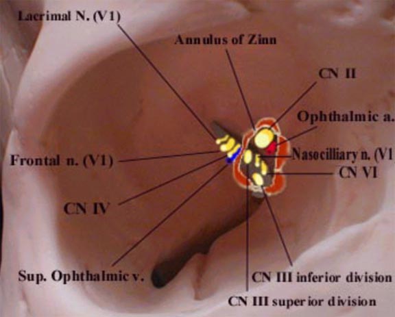
Relationship of nerves and vessels to the annulus of Zinn

Diagram of right orbit that shows the relationship of entering nerves and vessels to the annulus of Zinn.
 The four recti muscles arise from a short funnel-shaped tendinous ring called the annulus of Zinn which encloses the optic foramen and a part of the medial end of the superior orbital fissure. There are 2 tendons.
The Lower Tendon (of Zinn) is attached to the inferior root of the lesser wing of the sphenoid between the optic foramen and the superior orbital fissure. The lower tendon gives origin to part of the medial and lateral recti and all of the inferior rectus. The Upper Tendon (of Lockwood) arises from the body of the sphenoid, and gives origin to part of the medial and lateral recti and all of the superior rectus muscle. The superior and medial recti muscles are much more closely attached to the dural sheath of the optic nerve. This fact may be responsible for the characteristic pain which accompanies extreme eye movements in retro-bulbar neuritis.

Labels: , ,