Wednesday, December 29, 2010

Coronary Arterial Anatomy

Labels:

Tuesday, December 28, 2010

Brain Stem Model - Sagittal View - Thalamus & Epithalamus

Labels:

Monday, December 27, 2010

Layers of the Abdominal Wall with Function

The layers of the abdominal wall vary, depending on where it is you are looking.
At the lateral side of the abdomen there is a dotted line passing through the abdominal wall. Note the layers must pass through to .........

Labels:

Sunday, December 26, 2010

The lady between two majors


Bicipital groove: attachments of muscles near it

"The lady between two majors" :
Teres major attaches to medial lip of groove.
Pectoralis major to lateral lip of groove.
Latissimus (Lady) is on floor of groove, between the 2 majors.

Labels: ,

Thursday, December 23, 2010

Anatomy on Thyroid sonography

The anterior neck is depicted rather well with standard gray scale sonography. (FIGURE 1) The thyroid gland is slightly more echo-dense than the adjacent structures because of its iodine content. It has a homogenous ground glass appearance. Each lobe has a smooth globular-shaped contour and is no more than 3 - 4 centimeters in height, 1 - 1.5 cm in width, and 1 centimeter in depth. The isthmus is identified, anterior to the trachea as a uniform structure that is approximately 0.5 cm in height and 2 - 3 mm in depth.
The pyramidal lobe is not seen unless it is significantly enlarged. In the female, the upper pole of each thyroid lobe may be seen at the level of the thyroid cartilage, lower in the male. The surrounding muscles are of lower echogenicity than the thyroid and tissue planes between muscles are usually identifiable. The air-filled trachea does not transmit the ultrasound and only the anterior portion of the cartilaginous ring is represented by dense, bright echoes. The carotid artery and other blood vessels are echo-free unless they are calcified. The jugular vein is usually in a collapsed condition and it distends with a Valsalva maneuver. There are frequently 1-2 mm echo-free zones on the surface and within the thyroid gland that represent blood vessels. The vascular nature of all of these echoless areas can be demonstrated by color Doppler imaging to differentiate them from cystic structures (10-11).
Lymph nodes may be observed and nerves are generally not seen. The parathyroid glands are observed only when they are enlarged and are less dense ultrasonically than thyroid tissue because of the absence of iodine. The esophagus may be demonstrated behind the medial part of the left thyroid lobe, especially if it is distended by a sip of water. (FIGURE 2)
Figure 1. Sonogram of the neck in the transverse plane showing a normal right thyroid lobe and isthmus. L=small thyroid lobe in a patient who is taking suppressive amounts of L-thyroxine, I=isthmus, T=tracheal ring ( dense white arc is calcification, distal to it is artefact), C=carotid artery ( note the enhanced echoes deep to the fluid-filled blood vessel), J=jugular vein, S=Sternocleidomastoid muscle, m=strap muscle.

Figure 2. Sonogram of the left lobe of the thyroid gland in the transverse plane showing a rounded lobe of a goiter. L=enlarged lobe, I= widened isthmus, T=trachea, C=carotid artery ( note the enhanced echoes deep to the fluid-filled blood vessel), J=jugular vein, S=Sternocleidomastoid muscle, m=strap muscles, E=esophagus.

Labels: ,

Wednesday, December 22, 2010

Trachea and Esophagus in relation to vertebral and sternal levels

The trachea, or windpipe , which has cervical and thoracic parts, extends from the inferior end of the larynx (C6 vertebra) to its point of bifurcation (between T5 and 7 vertebral level). It is about 9 to 15 cm in length. .................

Labels:

Monday, December 20, 2010

Anatomy of aorta and appearance on x-ray


Labels: ,

Saturday, December 18, 2010

Basic surface anatomy knee

Labels: ,

Thursday, December 16, 2010

Anatomical parts of Gingiva

The image above shows the actual anatomy of the gingiva, known commonly as the "gums". The lighter pink colored gum tissue is called the "attached gingiva" because it is ..........

Labels: , ,

People have 3 types of tonsils

"PPL (people) have tonsils":
  • Pharyngeal
  • Palatine
  • Lingual

Labels: ,

Wednesday, December 15, 2010

Anatomy of ear drum as seen on examination

Clinical Anatomy: Applied Anatomy for Students and Junior Doctors 11th ed.

456 pages
Publisher: Wiley-Blackwell; 11 edition (November 17, 2006)
Language: English
ISBN-10: 1405138041
ISBN-13: 978-1405138048


Clinical Anatomy has remained the word of the same author for nearly half a century. Initially written for clinical medical students on the wards, clinics and revising for Finals, since then it has sold all over the world in hundreds of thousands and in several languages. Its success has been its constant revision to ensure it covers the student's needs. Today, it also covers the bulk of the anatomy required in the MRCS Part I, II, III and provides an excellent revision text for these candidates and medical students in a concise form with clear, easily reproduced diagrams.

For Download :

Labels: ,

Tuesday, December 14, 2010

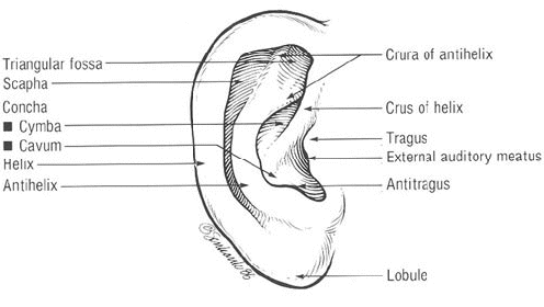
Surface Anatomy of the External Ear

The external ear consists of the auricle and the external auditory canal. The helix rim arises anteriorly and inferiorly from a crus extending horizontally above the external auditory meatus, thus............

Read more............ >>

Labels: , ,

Sunday, December 12, 2010

Anatomy on Radiographic Scapula

Labels: ,

Saturday, December 11, 2010

Popliteal fossa Mnemonic


Popliteal fossa: muscles arrangement

The two Semi's go together, Semimembranosus and Semitendonosus.
The Membranosus is Medial and since the two semis go together, Semitendonosus is also medial.

Therefore, Biceps Femoris has to be lateral.
Of the semi's, to remember which one is superficial: the Tendonosus is on Top.

Labels: ,

Friday, December 10, 2010

Fantastic Video about Auditory Transduction

This 7-minute video by Brandon Pletsch takes viewers on a step-by-step voyage through the inside of the ear, to the acoustic accompaniment of classical music.

Labels: , ,

Wednesday, December 8, 2010

Applied anatomy of the vital structures posterior to the sternoclavicular joint

Click on photo for enlarment

Applied anatomy of the vital structures posterior to the sternoclavicular joint. (A,B) Sagittal views in cross-section demonstrating the structures posterior to the sternoclavicular joint.
(C) A diagram demonstrating the close proximity of the major vessels posterior to the sternoclavicular joint.
(D) An aortogram showing the relationship of the medial end of the clavicle to the major vessels in the mediasternum.

Labels: ,

Relationship of nerves and vessels to the annulus of Zinn

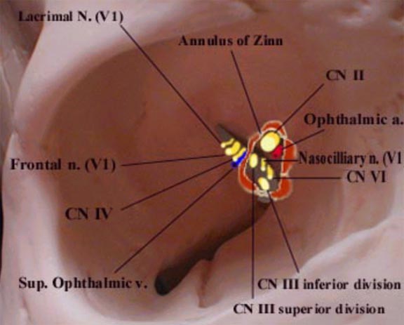
Diagram of right orbit that shows the relationship of entering nerves and vessels to the annulus of Zinn.
 The four recti muscles arise from a short funnel-shaped tendinous ring called the annulus of Zinn which encloses the optic foramen and a part of the medial end of the superior orbital fissure. There are 2 tendons.
The Lower Tendon (of Zinn) is attached to the inferior root of the lesser wing of the sphenoid between the optic foramen and the superior orbital fissure. The lower tendon gives origin to part of the medial and lateral recti and all of the inferior rectus. The Upper Tendon (of Lockwood) arises from the body of the sphenoid, and gives origin to part of the medial and lateral recti and all of the superior rectus muscle. The superior and medial recti muscles are much more closely attached to the dural sheath of the optic nerve. This fact may be responsible for the characteristic pain which accompanies extreme eye movements in retro-bulbar neuritis.

Labels: , ,

Tuesday, December 7, 2010

Anatomy, Histology, and Cell Biology PreTest Self-Assessment and Review, Third Edition (PreTest Basic Science)

This one-of-a-kind test prep guide helps you to test your knowledge of essential anatomy, histology and cell biology concepts for the USMLE Step 1; practice with 500 USMLE Step 1-style questions with referenced answers; review explanations for right and wrong answers;and build confidence, skills, and knowledge.

Labels: , ,

Anatomical Considerations of Abdominal aorta in Ultrasound

The abdominal aorta is entirely a retroperitoneal structure. It begins at the aortic hiatus of the diaphragm at approximately the 12th thoracic vertebrae and courses anterior to the spine before bifurcating into the common iliac arteries. The inferior vena cava (IVC) courses to the right of the abdominal aorta and may be confused for the abdominal aorta by novice sonologists. The psoas muscle and kidney are posterior-lateral to the abdominal aorta on the left side............

Read more..............>>

Branches of the abdominal aorta and inferior vena cava. SMA = superior mesenteric artery.......

Read more..................>>

Labels: ,