Tuesday, January 4, 2011

Le Fort Classification of facial fractures

In 1901 ;René Le Fort (1869-1951),French surgeon, reported his work on cadaver skulls that were subjected to blunt forces of various magnitudes and directions. He concluded that predictable patterns of fractures follow certain types of injuries. Three predominant types were described.


Le Fort I fractures (horizontal) may result from a force of injury directed low on the maxillary alveolar rim in a downward direction. The fracture extends from the nasal septum to the lateral pyriform rims, travels horizontally above the teeth apices, crosses below the zygomaticomaxillary junction, and traverses the pterygomaxillary junction to interrupt the pterygoid plates.

Le Fort II fractures (pyramidal) may result from a blow to the lower or mid maxilla. Such a fracture has a pyramidal shape and extends from the nasal bridge at or below the nasofrontal suture through the frontal processes of the maxilla, inferolaterally through the lacrimal bones and inferior orbital floor and rim through or near the inferior orbital foramen, and inferiorly through the anterior wall of the maxillary sinus; it then travels under the zygoma, across the pterygomaxillary fissure, and through the pterygoid plates.

Le Fort III fractures (transverse), also termed craniofacial dysjunctions, may follow impact to the nasal bridge or upper maxilla. These fractures start at the nasofrontal and frontomaxillary sutures and extend posteriorly along the medial wall of the orbit through the nasolacrimal groove and ethmoid bones. The thicker sphenoid bone posteriorly usually prevents continuation of the fracture into the optic canal. Instead, the fracture continues along the floor of the orbit along the inferior orbital fissure and continues superolaterally through the lateral orbital wall, through the zygomaticofrontal junction and the zygomatic arch. Intranasally, a branch of the fracture extends through the base of the perpendicular plate of the ethmoid, through the vomer, and through the interface of the pterygoid plates to the base of the sphenoid.

Labels: ,

Monday, January 3, 2011

The staging evaluation of Esophageal Cancer

After establishing a diagnosis of esophageal cancer, adequate staging is required, because staging is the most important step in choosing appropriate therapy. More than 50% of patients have unresectable or metastatic disease at the time of presentation. For the others, survival is closely related to the stage of the disease.
The staging evaluation allows patients to be ...................

Labels: ,

Sunday, January 2, 2011

Diagram for Effects of sickle cell anemia

Monday, December 27, 2010

Complications of Sickle cell disease

Medical mnemonic for the Sickle cell disease complications

SICKLE:

Strokes/ Swelling of hands and feet/ Spleen problems
Infections/ Infarctions
Crises (painful, sequestration, aplastic)/ Cholelithiasis/ Chest syndrome/ Chronic hemolysis/ Cardiac problems
Kidney disease
Liver disease/ Lung problems
 Erection (priapism)/ Eye problems (retinopathy)

Labels: , ,

Sunday, December 26, 2010

Synthesis of eicosanoid autacoids

The eicosanoids are an important group of endogenous fatty acid derivatives that are produced from arachidonic acid, a 20-carbon fatty acid lipid in cell membranes................

Labels: , ,

Diagram of meniscal tear patterns

Meniscal injuries can be classified according to their tear patterns. A vertical or longitudinal tear occurs in line with the circumferential fibers of the meniscus . If long enough, this tear is known as a bucket-handle tear. At arthroscopy, the bucket-handle tear may be seen as being attached anteriorly and posteriorly. Alternatively, it may be detached at either end or transected in the middle with unstable anterior and posterior flaps. A bucket-handle tear may displace into the intercondylar notch, where it may cause true locking of the knee joint.
(A) Vertical or longitudinal (Bucket-handle), (B) Flap or Oblique, (C) Radial or Transverse, (D) Horizontal, (E) Complex degenerative.

-Oblique tears are also known as flap or parrot beak tears and are perhaps the most common . These occur generally at the junction of the posterior and middle thirds.

-Radial tears occur in a similar location. They extend from the inner free margin toward the periphery . If such a tear reaches the periphery, it transects the meniscus and renders the hoop stress-distributing capacities of the meniscus useless. Such a tear is the functional equivalent of a total meniscectomy.

-Horizontal cleavage tears usually occur in older individuals. They extend from the inner free margin peripherally to the intrameniscal substance where myxoid degeneration may be present. These tears divide the meniscus into superior and inferior flaps, either of which may be unstable

-Complex degenerative tears occur in older patients. Osteoarthritic changes may be visible on plain radiographs, and chondromalacia of the articular surfaces is commonly encountered. The tears occur in multiple planes .

Labels: ,

Saturday, December 25, 2010

Classification of inflammatory arthritis

Inflammatory arthritis is generally classified into seropositive and seronegative groups. These are based on the presence of rheumatoid factor, an immunoglobulin which reacts with gamma globulin, in the blood of the majority of patients with seropositive disease and in a small minority of patients with seronegative disease. The prototype seropositive form of arthritis is rheumatoid arthritis. Other members include the group of conditions labelled collagen vascular diseases, such as systemic lupus erythematosus, scleroderma, vasculitis, Sjogren’s syndrome. Only rheumatoid arthritis will be considered in this paper.

Among the seronegative inflammatory joint diseases is a group labelled spondyloarthritis. This condition is characterized by inflammatory disease of the joints of the back, both the sacroiliac joints and the apophyseal joints of the spine. Members of this group include ankylosing spondylitis, psoriatic arthritis, reactive arthritis, and arthritis of inflammatory bowel disease. While the prototype for this group is ankylosing spondylitis, psoriatic arthritis will also be discussed.

In addition to the presence of rheumatoid factor, there are extra-articular features which distinguish the seropositive from the seronegative forms of inflammatory arthritis.

Labels: ,

Saturday, December 18, 2010

Hyper Vs Hypothyroidism

Labels: ,

Thursday, December 16, 2010

Shapes of Arterial pressure waves

While palpating the pulse of a patient, you note that the pulse wave has two peaks. You auscultate the heart and are certain that there is only one heartbeat for each two
pulse waves. Which of the following best describes this finding?
  1. a.Pulsus alternans
  2. b.Dicrotic pulse
  3. c.Pulsus parvus et tardus
  4. d.Pulsus bigeminus
  5. e.Pulsus bisferiens

. The answer is: ( e ).
 Pulsus bisferiens (bisferious pulse) is seen in AI and in hypertrophic cardiomyopathy (HCM). In the latter, the first wave or percussion wave is due to the rapid flow rate of initial contraction, and the second wave or tidal wave is due to the slower rate of continued contraction. The dicrotic pulse has two palpable pulses, but one is in systole and the other is in diastole.

Pulsus bigeminus is an alteration in pulse amplitude that follows a ventricular premature beat. Pulsus alternans is a regular alternating pulse amplitude due to alternating left ventricular contractile force; it is usually seen with severe left ventricular decompensation and cardiac tamponade. Pulsus parvus et tardus(“small and slow rising”) represents a delayed systolic peak due to obstruction to left ventricular ejection. It is seen in aortic stenosis (AS)


CLICK FOR MORE DETAILES

Labels: , ,

Wednesday, December 15, 2010

Anatomy of ear drum as seen on examination

The Confusion Assessment Model (CAM)


CLICK for enlarment

Labels: , ,

Tuesday, December 14, 2010

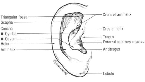
Surface Anatomy of the External Ear

The external ear consists of the auricle and the external auditory canal. The helix rim arises anteriorly and inferiorly from a crus extending horizontally above the external auditory meatus, thus............

Read more............ >>

Labels: , ,

Thursday, December 9, 2010

Diagrams for Surgical Procedures of Chronic Pancreatitis

The selection of the operative procedure often depends on the surgeon's expertise and individual preference. There is some criteria that usually favor one type of surgery over another. When the pancreatic duct in the body or tail is dilated beyond 6 mm, the Puestow procedure is usually the most effective surgery . When disease occurs predominantly in the head, Frey's procedure is used. When there is a focal mass in the head without significant duct dilation, the Whipple procedure is most frequently used. Increasingly, Beger's procedure, which preserves the duodenum, is used as an alternative.

1-Whipple Procedure:
The most common pancreatic resection surgery is the Whipple procedure, which is performed for chronic pancreatitis with ductal strictures or amorphous inflammatory mass in the head for which distinction from cancer cannot be made preoperatively. However, many specialist pancreatic surgeons no longer consider this to be the most appropriate surgery for chronic pancreatitis.
Diagram of pylorus-preserving Whipple procedure. Classic Whipple procedure is shown in inset: It entails radical dissection of pancreatic head, adjacent nodes, right half of omentum, gall bladder, common bile duct, and most or all of duodenum followed by gastrojejunostomy/duodenojejunostomy (green arrow), pancreaticojejunostomy (blue arrow), and hepaticojejunostomy (red arrow). (Courtesy of the Office of Visual Media, Indiana University)

2-Beger's Procedure :
Beger's procedure is a less radical surgery with resection of the pancreatic head and preservation of the duodenum . After Beger's surgery, pain relief is seen in up to 85% of patients at 5-year follow-up , but the postoperative morbidity rate is 20%. A randomized study comparing Beger's and Whipple procedures showed similar results at 6 months except for better pain tolerance and glucose control in those treated with Beger's surgery.
Diagram of Beger's procedure. Pancreatic body and most of head have been resected. Sleeve of pancreas is left with duodenum to preserve blood supply for latter. This procedure is technically harder to perform than Whipple procedure. Note pancreaticojejunostomy (red arrows) at two sites of Roux limb (green arrow).

3-Puestow Procedure :
The Puestow procedure is a side-to-side longitudinal pancreaticojejunostomy that drains the pancreatic duct directly into a loop of jejunum . This procedure is best performed if the main pancreatic duct is significantly dilated, usually wider than 6 mm.
Diagram of Puestow procedure. Pancreas is filleted to expose main duct from neck to tail and ductal calculi are removed. Roux loop is anastomosed to "capsule" of pancreas with direct drainage of main and secondary pancreatic ducts into jejunum over 8- to 10-cm segment. Loop (arrows) lies anterior to pancreas.

4-Frey's Procedure :
Frey's procedure is a recently popularized procedure that combines partial resection of the pancreatic head with a longitudinal jejunostomy . The morbidity rate of Frey's procedure is approximately 9-22% , well below that of the Whipple procedure performed for chronic pancreatitis, for which the complication rate is 30-40% . Frey's procedure is contraindicated in the presence of duodenal or biliary stricture.
Diagram of pancreas after Frey's procedure. Head of pancreas is cored out (blue arrow) and pancreaticojejunostomy is created via Roux loop (green arrows). Procedure is best performed in patients with duct dilation of head and body.

Labels: ,

Wednesday, December 8, 2010

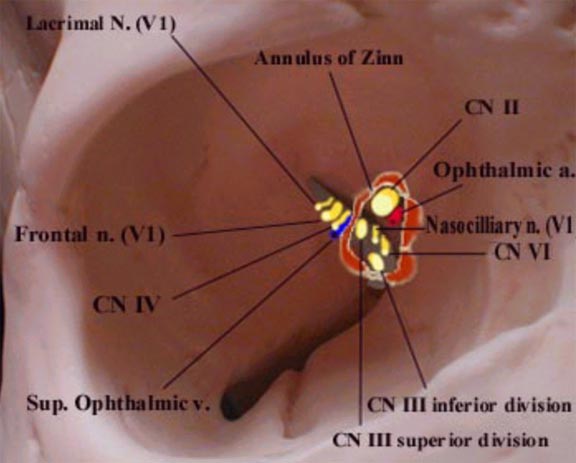
Relationship of nerves and vessels to the annulus of Zinn

Diagram of right orbit that shows the relationship of entering nerves and vessels to the annulus of Zinn.
 The four recti muscles arise from a short funnel-shaped tendinous ring called the annulus of Zinn which encloses the optic foramen and a part of the medial end of the superior orbital fissure. There are 2 tendons.
The Lower Tendon (of Zinn) is attached to the inferior root of the lesser wing of the sphenoid between the optic foramen and the superior orbital fissure. The lower tendon gives origin to part of the medial and lateral recti and all of the inferior rectus. The Upper Tendon (of Lockwood) arises from the body of the sphenoid, and gives origin to part of the medial and lateral recti and all of the superior rectus muscle. The superior and medial recti muscles are much more closely attached to the dural sheath of the optic nerve. This fact may be responsible for the characteristic pain which accompanies extreme eye movements in retro-bulbar neuritis.

Labels: , ,

Sunday, December 5, 2010

Lumbrical action

Lumbrical action is to hold a pea, that is to flex the metacarpophalangeal joint and extend the interphalangeal joints. When look at hand in this position, can see this makes an “L” shape, since L is for Lumbrical...............

Read more..........>>

Labels: ,

Saturday, November 27, 2010

Mechanisms Linked to Sodium intake in Hypertension

Mechanisms Linked to Increases in Blood Pressure and the Therapeutic Effects of Healthful Dietary Patterns, Sodium Reduction, and Weight Loss.

Sodium intake initiates an autoregulatory sequence that leads to increased intravascular fluid volume and cardiac output, peripheral resistance, and blood pressure. The elevation in blood pressure results in a phenomenon called pressure natriuresis, in which increased renal perfusion pressure leads to increased excretion of fluid and sodium. In essential hypertension, however, sodium excretion is impaired. It is hypothesized that in most cases essential hypertension is a genetic disorder involving many individual genes, each of which influences the body's handling of sodium to varying degrees and becomes expressed in the context of an unhealthful dietary environment, particularly one characterized by excessive intake of salt.

Labels: , ,

Wednesday, November 24, 2010

RIGHT VENTRICULAR FAILURE IN COPD


Severe pulmonary hypertension increases right ventricular afterload and eventually leads to the clinical syndrome of right heart failure with systemic congestion and inability to adapt right ventricular output to peripheral demand at exercise. Many patients with advanced COPD present with ankle edema but normal right atrial pressures (at rest). This apparent paradox has stimulated speculation that edema in COPD might be a renal rather than a right ventricular problem .


However, it is now better realized that edema in COPD is likely to be initially caused by repeated stretching of the right atrium from increased right ventricular diastolic pressures at exercise or conceivably with oxygen desaturation during sleep, causing increased sympathetic nervous system tone and activation of the renin-angiotensin-aldosterone system, with resultant renal salt and water retention.
 Renal salt and water retention may be aggravated by hypercapnia, which directly increases proximal tubular reabsorption of sodium, but also activates the sympathetic nervous system and the renin-angiotensin-aldosterone system, which causes additional distal tubular sodium reabsorption through amiloride-sensitive sodium channels.

In summary, and as summarized in , systemic congestion in COPD is caused by right heart failure, involving mechanisms that are very similar to those accounting for systemic and pulmonary congestion in left heart failure, but with an important additional contribution of hypercapnia .

Labels: , ,

Friday, November 19, 2010

Glasgow Coma Scale

A 29-year-old woman was an unbelted passenger in a motor vehicle accident. On arrival to the hospital, the paramedics inform you that she opens her eyes in response to verbal stimuli. She is incoherent and withdraws from painful stimuli. Which of the following is the patient’s calculated Glasgow Coma Scale (GCS)?
  • a.15
  • b.3
  • c.9
  • d.5
  • e.12

The answer is (C). The Glasgow Coma Scale (GCS) is often used to quantify consciousness and assess cerebral cortex and brain stem function by assessing the patient’s verbal response, motor response, and eye opening response to stimuli.
It may be repeated at intervals to detect improvement or deterioration and is now widely used in coma assessment. The minimum score is 3 and the maximum score is 15.
Three behaviors are assessed in the GCS:

Labels: , ,

Friday, November 5, 2010

Lymphatic Drainage in Axillary Lymph Nodes

The lymphatic vessels of the upper limb, most of those from the breast, and the cutaneous vessels of the trunk above the level of the umbilicus drain into the axillary nodes.

Lymphatics from the fingers accompany the cephalic and basilic veins and enter the lateral axillary and deltopectoral (or infraclavicular) nodes .

Axillary Nodes. These important nodes are arbitrarily divided into five groups:
1. The lateral nodes lie behind the axillary vein and drain the upper limb.

2. The pectoral nodes, at the inferior border of the pectoralis minor, drain most of the breast.

3. The posterior, or subscapular, nodes, in the posterior axillary fold, drain the posterior shoulder.

4. The central nodes, near the base of the axilla, receive the lymph from the preceding three groups. They form the group most likely to be palpable (against the lateral thoracic wall).

5. The apical nodes lie medial to the axillary vein and superior to the pectoralis minor. The apical nodes receive the lymph from all the other groups and sometimes directly from the breast. They drain into two or three subclavian trunks, which enter the jugular-subclavian venous confluence, or join a common lymphatic duct, or empty into lower, deep cervical nodes.
Diagram of the lymphatic drainage of the upper limb and breast. The supratrochlear and deltopectoral nodes receive many superficial lymphatic vessels. The axillary nodes are indicated by capital letters. The lateral nodes drain the upper limb. The subareolar plexus drains by collecting trunks into the axillary nodes. The pectoral nodes drain most of the breast. The apical nodes receive the lymph from the other axillary groups. Retropectoral (R) and transpectoral (T) routes are also shown.

Labels: ,

Sunday, October 31, 2010

Surface anatomy of the Stomach

The cardiac orifice is opposite the 7th left costal cartilage about 2.5 cm. from the side of the sternum; it corresponds to the level of the tenth thoracic vertebra.
The pyloric orifice is on the transpyloric line about 1 cm. to the right of the middle line, or alternately 5 cm. below the 7th right sternocostal articulation; it is at the level of the first lumbar vertebra.
A curved line, convex downward and to the left, joining these points indicates the lesser curvature. In the left lateral line the fundus of the stomach reaches as high as the 5th interspace or the 6th costal cartilage, a little below the apex of the heart.

To indicate the greater curvature a curved line is drawn from the cardiac orifice to the summit of the fundus, thence downward and to the left, finally turning medialward to the pyloric orifice, but passing, on its way, through the intersection of the left lateral with the transpyloric line. The portion of the stomach which is in contact with the abdominal wall can be represented roughly by a triangular area the base of which is formed by a line drawn from the tip of the 10th left costal cartilage to the tip of the 9th right cartilage, and the sides by two lines drawn from the end of the 8th left costal cartilage to the ends of the base line.

Note That: This measurements given refer to a moderately filled stomach with the body in the supine position !!!  Why we say that.........
As The shape of the stomach is constantly undergoing alteration; it is affected by the particular phase of the process of gastric digestion, by the state of the surrounding viscera, and by the amount and character of its contents. Its position also varies with that of the body.
With the patient in the erect posture.
With the patient lying down.

Labels: ,