Friday, January 7, 2011

How to appreciate Traumatic exophthalmos

Traumatic exophthalmos develops as a retrobulbar hematoma that pushes the eyeball globe outward. Patient presents with periorbital edema, ecchymosis, a marked decrease in visual acuity, and an afferent pupillary defect in the involved eye.
Sometimes the exophthalmos may be obscured by ..........

Read more.........>>

Labels: ,

Tuesday, January 4, 2011

ABC of Resuscitation (ABC series)

111 pages
Publisher: BMJ Books; 5 edition (January 7, 2004)
Language: English
ISBN-10: 0727916696
ISBN-13: 978-0727916693


Product Description
This authoritative guide has concise and practical information on all aspects of resuscitation. New guidelines are just one of the changes to the 5th edition of this book as many of the chapters have been completely rewritten,with new material on automated defibrillators, advanced life support including paediatric life support, new drugs plus many new illustrations. . The text has worldwide relevance with particular emphasis on Europe, Australasia, South Africa, South America, Malaysia and the Middle East.

For Download :

Labels: ,

Sunday, January 2, 2011

Pneumocephalus

X-ray showing nasal and orbital fractures and pneumocephalus
Pneumocephalus is the presence of air in the cranial vault. It is usually associated with neurosurgery, barotrauma, basilar skull fractures, sinus fractures, nasopharyngeal tumor invasion and meningitis..........

Labels: ,

Thursday, December 30, 2010

Procedures to open obstructed airway

#The tongue is the single most common cause of an airway obstruction. In most cases, the airway can be cleared by simply using the head-tilt/chin-lift technique. This action pulls the tongue away from the air passage in the throat ,See this video:
Head tilt chin lift Technique


#The head-tilt/chin-lift is an important procedure in opening the airway; however, use extreme care because excess force in performing this maneuver may cause further spinal injury. In a casualty with a suspected neck injury or severe head trauma, the safest approach to opening the airway is the jaw-thrust technique because in most cases it can be accomplished without extending the neck.See it:
Jaw Thrust Technique

Labels: ,

Wednesday, December 29, 2010

Assessment of Head injuries in children

Perform a primary survey and ensure that the child’s airway, cervical spine, breathing and circulation are secure.

Rapidly assess the child’s mental state using the AVPU scale. Use firm supraorbital pressure as the painful stimulus.

* A Alert
* V Responds to voice
* P Responds to pain
----> Purposefully
----> Non-purposefully :
  •  Withdrawal/flexor response
  •  Extensor response
* U Unresponsive
Assess pupil size, equality and reactivity and look for other focal neurological signs.
Perform a secondary survey looking specifically at:

* Neck and cervical spine – deformity, tenderness, muscle spasm
* Head – scalp bruising, lacerations, swelling, tenderness, bruising behind the ear (Battles sign)
* Eyes – pupil size, equality and reactivity, fundoscopy
* Ears – blood behind the ear drum, CSF leak
* Nose – deformity, swelling, bleeding, CSF leak
* Mouth –dental trauma, soft tissue injuries
* Facial fractures
* Motor function – examine limbs for presence of reflexes and any lateralising weakness
* Perform a formal Glasgow Coma Score
* Consider the possibility of non-accidental injury during secondary survey especially in infants with head injury.
* Other injuries (see major trauma guidelines)

Labels: , ,

Photos for Battle's sign


Battle's sign, also called mastoid ecchymosis : consists of bruising over the mastoid process (just behind the auricle), as a result of extravasation of blood along the path of the posterior auricular artery.

It is an indication of fracture of the base of the posterior portion of the skull, and may suggest underlying brain trauma.

Labels: ,

Brain herniation Sites

Brain herniation refers to displacement of a portion of the brain from its normal position through openings in the inelastic dura secondary to.............

Read more.............>>

Labels: ,

Tuesday, December 28, 2010

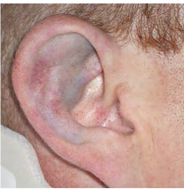
Pinna Contusion and Hematoma

Blunt external ear trauma may cause a contusion or hematoma of the pinna which, if untreated, may result in cartilage necrosis and chronic scarring or further cartilage formation and permanent deformity or "cauliflower ear." Open injuries include lacerations (with and without cartilage exposure) and avulsions.
Pinna Contusion. Contusion without hematoma is present...........


Labels: ,

Monday, December 27, 2010

The Atlas of Emergency Medicine 3rd edition


The most complete and trusted visual compendium of emergency medicine—extensively updated with 1500 full-color illustrations
Ideally suited to the bedside practice of emergency medicine, The Atlas of Emergency Medicine is the ultimate visual guide to the diagnosis and treatment of common and uncommon conditions encountered in the Emergency Department. Filled with 1500 crisp, clear full-color images, this essential clinical companion is logically organized by organ system and then by problem, making it a practical quick reference for medical students, residents in training, new graduates preparing for their certification exam, the practicing physician, and instructors. The third edition of The Atlas of Emergency Medicine features an even more streamlined presentation with clear, concise text and an unmatched collection of diagnoses-speeding images. Forming the core of the book, these images show you what to look for and are accompanied by brief, high-yield descriptions of clinical problems. The new edition also features an enhanced template, and new coverage of airway emergencies, tropical conditions, toxicologic emergencies, and electrocardiographs.
NEW TO THIS EDITION
* 1,500 full-color clinical photographs (more than twice the amount found in the previous edition)
New chapter template:“Clinical Summary”: clinically relevant observations on differential diagnosis
“Emergency Department Treatment and Disposition”: a brief overview of need-to-know diagnostic guidelines and recommendations
“Clinical Pearls”: instructive tips and insights on specific aspects of conditions which are difficult to find in other texts
*Four new chapters that enhance the book’s hands-on value:
Tropical Medicine: reflects an increased emphasis on global access to healthcare and easier patient travel
Toxicology: features an up-to-date, authoritative review of how to diagnose and treat selected toxicological emergencies
ECG Abnormalities: presents turnkey insights into the rapid recognition of pathological ECG’s, highlighting the electrocardiographic characteristics of each featured clinical problem
Airway Procedures: includes essential information that guides, improves, and expedites the management of airway emergencies

For Download :

Labels: ,

Thursday, December 16, 2010

BLUNT ABDOMINAL TRAUMA(Surgery algorithm)

Wednesday, December 15, 2010

The Confusion Assessment Model (CAM)


CLICK for enlarment

Labels: , ,

Sunday, December 5, 2010

Button battery in ear; How dangerous is it ?

Young children commonly place foreign bodies in the ear canal and nose. If a vegetable foreign body is present, do not use ear drops because they will cause the foreign body to swell.

One of the most dangerous foreign bodies is a button battery. This type of foreign body should be removed immediately. Prior to removal, the use of ear drops of any kind is absolutely contraindicated.
The electrical charge of the battery will produce electrolysis of any electrolyte-rich fluid. This produces hydroxides which will cause a severe alkaline burn. The picture above shows a severe burn of the external auditory canal which resulted in the exposure of bone over 180 degrees of the ear canal's circumference. The battery was in the child's ear for approximately three days and the picture was taken one month after the battery was removed. Healing is slow and damage to the eardrum, facial nerve, and inner ear can occur. Luckily in this patient the injury was confined to the ear canal.

Labels: , ,

Tuesday, November 30, 2010

Appearance of lye strictures in X-ray


-What is the most likely diagnosis in the patient shown below?

a. Esophageal varices
b. Esophageal carcinoma
c. Reflux esophagitis
d. Lye stricture
e. Fibrovascular polyp


THE RIGHT ANSEWR :
.d

There is very long segment of smoothly narrowed esophagus from just below the level of the clavicles to the EG junction. This appearance is characteristic ............

Labels: , , ,

Monday, November 29, 2010

Ultrasound use in trauma - the fast exam

instructional video describing ultrasound use in trauma - the fast exam

Labels: ,

Sunday, November 28, 2010

Ileocecal Intussusception

A 45-year-old man with no notable medical or surgical history presented with a 24-hour history of intense pain in the right side of the abdomen with associated nausea and vomiting. He reported having had similar but much less severe episodes during the previous 6 months. Results of initial laboratory tests were unrevealing.
Physical examination showed moderate abdominal distention. Computed tomographic scans of his abdomen revealed an ileocecal intussusception (Panel A, arrow) with a pathologic mass, 2.5 cm in diameter, at the apex, also known as the lead point (Panel B, arrow). Diagnostic laparoscopy was performed, and the diagnosis of intussusception was confirmed.

Laparoscopically assisted ileocecal resection with primary anastomosis was performed. Gross inspection of the specimen showed a pedunculated lipoma within the terminal ileum. The patient had a rapid recovery, with complete resolution of his symptoms.

Labels: , , ,

Wednesday, November 24, 2010

Yearbook of Intensive Care and Emergency Medicine - Febuary 2010 Edition


The Yearbook compiles the most recent developments in experimental and clinical research and practice in one comprehensive reference book. The chapters are written by well recognized experts in the field of intensive care and emergency medicine. It is addressed to everyone involved in internal medicine, anesthesia, surgery, pediatrics, intensive care and emergency medicine.

Labels: , ,

Saturday, November 20, 2010

causes of coma, mnemonic

There are several medical causes of coma. One of the easiest ways to remember these causes is to use the mnemonic AEIOU TIPS:

* A - alcohol (overdose / withdraw / Wernicke's encephalopathy)
* E - epilepsy or exposure (heat stroke, hypothermia) or electrolytes
* I - insulin (diabetic emergency - high/low sugar)
* O - overdose or oxygen deficiency
* U - uremia (metabolic) or underdose
* T - trauma (head injury) or toxicology
* I - infection / inflammation
* P - psychosis or poisoning
* S - stroke / SAH / shock / syncope

Labels: , ,

Friday, November 19, 2010

Glasgow Coma Scale

A 29-year-old woman was an unbelted passenger in a motor vehicle accident. On arrival to the hospital, the paramedics inform you that she opens her eyes in response to verbal stimuli. She is incoherent and withdraws from painful stimuli. Which of the following is the patient’s calculated Glasgow Coma Scale (GCS)?
  • a.15
  • b.3
  • c.9
  • d.5
  • e.12

The answer is (C). The Glasgow Coma Scale (GCS) is often used to quantify consciousness and assess cerebral cortex and brain stem function by assessing the patient’s verbal response, motor response, and eye opening response to stimuli.
It may be repeated at intervals to detect improvement or deterioration and is now widely used in coma assessment. The minimum score is 3 and the maximum score is 15.
Three behaviors are assessed in the GCS:

Labels: , ,

Wednesday, November 3, 2010

Algorithm for treatment of renal trauma

The Liver Injury Scale (LIS) classification

The liver is the largest solid abdominal organ with a relatively fixed position, which makes it prone to injury . Damage to the liver is the most common of death after abdominal injury. The most common cause of liver injury is blunt abdominal trauma, which is secondary to motor vehicle crashes (MVC)

Hepatic injury was graded according to the Hepatic Injury Scale established by the American Association for the Surgery of Trauma (AAST).
This is a liver injury . The injury has been opened to control bleeding branches of the portal and hepatic veins as well as the hepatic arterial radicles. Several biliary ducts were ligated.


Grade and Description of injury
 Grade I : *Haematoma--->Subcapsular, non-expanding, less than 10 percent of surface area
*Laceration--->Capsular tear, non-bleeding, parenchymal depth less than 1 cm

Grade II : *Haematoma--->Subcapsular, non-expanding, 10–50 per cent of surface area or intraparenchymal, non-expanding, less than 2 cm in diameter.
*Laceration--->Capsular tear, active bleeding, parenchymal depth 1–3 cm, less than 10 cm in length.

Grade III : *Haematoma--->Subcapsular, more than 50 per cent of surface area or expanding ruptured subcapsular haematoma with active bleeding intraparenchymal haematoma larger than 2 cm.
*Laceration--->Parenchymal depth more than 3 cm.

Grade VI : *Haematoma--->Ruptured intraparenchymal haematoma with active bleeding.
*Laceration--->Parenchymal disruption of more than 25–50 percent of hepatic lobe.

Grade V : *Laceration--->Parenchymal disruption of more than 50 per cent of hepatic lobe.
*Vascular--->Juxtahepatic venous injuries.

Grade VI : *Vascular--->Hepatic avulsion.

Labels: ,