Tuesday, January 4, 2011

Synthesis & Transport of Thyroid Hormones

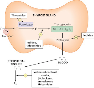
The thyroid secretes 2 iodine-containing hormones: thyroxine (T4 ) and triiodothyronine (T3). The iodine necessary for the synthesis of these molecules comes from food or iodide supplements. Iodide ion is actively taken up by and highly concentrated in the thyroid gland, where it is converted to elemental iodine by thyroidal peroxidase ( See the Figure ).
The protein thyroglobulin serves as a scaffold for thyroid hormone synthesis. Tyrosine residues in thyroglobulin are iodinated to form monoiodotyrosine (MIT) or diiodotyrosine (DIT) in a process known as iodineorganification........

Read more..........>>

This figure also show Sites of action of some antithyroid drugs. I–, iodide ion; I°, elemental iodine. Not shown: radioactive iodine (131I), which destroys the gland through radiation.

Labels: ,

Thursday, December 30, 2010

GOUT PIC


Gout of the Right Great Toe: Diffuse swelling and redness centered at the right MTP joint, but extending
over much of the foot.


Gout of the Left Great Toe: Diffuse swelling and redness centered at the left MTP joint.

Labels: ,

Thursday, December 23, 2010

Osteoporosis of aging (senile or postmenopausal osteoporosis)

Most common form of generalized osteoporosis. As a person ages, the bones lose density and become more brittle, fracturing more easily and healing more slowly. Many elderly persons are also less active and have poor diets that are deficient in protein. Females are affected more often and more severely than males, as postmenopausal women have deficient gonadal hormone levels and decreased osteoblastic activity.

 Osteoporosis of aging. Generalized demineralization of the spine in a postmenopausal woman. The cortex appears as a thin line that is relatively dense and prominent (picture-frame pattern).

Labels: , ,

Wednesday, December 22, 2010

Scheme for Metabolic acidosis

Metabolic acidosis is a commonly presenting feature and is often caused by diabetes, renal failure or poisoning. However, it can cause diagnostic difficulties, particularly in the acute situation when patients ................

Labels: ,

Sunday, December 19, 2010

Screening for Diabetic Retinopathy by new Fundus Camera

Saturday, December 18, 2010

Hyper Vs Hypothyroidism

Labels: ,

Thursday, December 16, 2010

Eliciting Chvostek’s sign

Chvostek's sign is contraction of the muscles of the eye, mouth or nose, elicited by tapping along the course of the facial nerve. The examiner taps gently over the facial nerve in front of the ear.

This sign usually suggests hypocalcemia but can occur normally in about 25% of patients. Typically, it precedes other signs of hypocalcemia and persists until the onset of tetany. It can’t be elicited during tetany because of strong muscle contractions.
Normally, eliciting Chvostek's sign is attempted only in patients with suspected hypocalcemic disorders. However, because the parathyroid gland regulates calcium balance, Chvostek's sign may also be tested in patients before neck surgery to obtain a baseline.

Labels: ,

Tuesday, November 23, 2010

Male Reproductive System: Hormone Pathways

A lecture on the hormones that play a role in the male reproductive system, with emphasis on the hormonal pathways.

Labels: ,

Sunday, November 21, 2010

USMLE ALGORITHMS: Cushing Syndrome

This video explains the complete workup of Cushing Syndrome, the causes, the diagnosis, and the full management. It is very thorough. I hope you enjoy

Labels: ,

Saturday, November 20, 2010

Trousseau sign of latent tetany

Trousseau’s sign presents as carpopedal spasm occurring after a few minutes of inflation of a sphygmomanometer cuff above ............

Labels: , , ,

Wednesday, November 17, 2010

Achondroplasia


Achondroplasia is an inherited disorder of bone growth. It is one of the group of disorders that are collectively called chondrodystrophies or osteochondrodysplasias.

The disorder causes a type of dwarfism that is............

Labels: ,

Monday, November 15, 2010

Clinical Approach to Metabolic Alkalosis

History
Obtain historical data to pinpoint the nature of the disease causing metabolic alkalosis.

* Ask the patient about history of vomiting, other gastric fluid loss, and diuretic use. Loss of gastric fluid and HCl due to ..............

Read more...........>>


Labels: , ,

Friday, November 5, 2010

Surgical Debridement of an Infected Diabetic Foot Wound

Thursday, November 4, 2010

Disease of bones,stones,abdominal groans and psychiatric moans

Primary hyperparathyroidism is described as "a common disorder of mineral metabolism characterized by incompletely regulated, excessive secretion of parathyroid hormone from one or more of the parathyroid glands"

The signs and symptoms of primary hyperparathyroidism are those of hypercalcemia. They are classically summarized by the mnemonic "stones, bones, abdominal groans and psychiatric moans".

* "Stones" refers to kidney stones, nephrocalcinosis, and diabetes insipidus (polyuria and polydipsia). These can ultimately lead to renal failure.
* "Bones" refers to bone-related complications. The classic bone disease in hyperparathyroidism is osteitis fibrosa cystica, which results in pain and sometimes pathological fractures. Other bone diseases associated with hyperparathyroidism are osteoporosis, osteomalacia, and arthritis.
* "Abdominal groans" refers to gastrointestinal symptoms of constipation, indigestion, nausea and vomiting. Hypercalcemia can lead to peptic ulcers and acute pancreatitis.
* "Psychiatric moans" refers to effects on the central nervous system. Symptoms include lethargy, fatigue, depression, memory loss, psychosis, ataxia, delirium, and coma.

The German description of the same symptoms is "Stein-, Bein- und Magenpein", literally "stone, leg, and stomach-pain".

In 1990, A National Institutes of Health (NIH) consensus panel defined renal stones in patients with primary hyperparathyroidism as an absolute indication for parathyroidectomy.
2-year-old woman  underwent parathyroidectomy for primary hyperparathyroidism. Sonogram of left kidney shows multiple calculi (arrows).

Labels: , ,

Tuesday, November 2, 2010

Diabetes-Related Atherosclerosis

Thursday, October 28, 2010

Acromegaly

Acromegaly 1

Acromegaly 2
Facial changes secondary to elevated growth hormone levels. Note in particular
prominent supra-orbital ridge, jaw, and generally enlarged facial features.

FOR MORE IFORMATION:
CLICK HERE

Labels: ,

Tuesday, October 26, 2010

Paget's disease of the Mandible


A 55-year-old man presented with a 2-year history of painful jaw enlargement and progressively ill-fitting dentures. He had no headaches or visual-field defects and did not have hyperhidrosis, oily skin, glucose intolerance, heart failure, or an increase in glove or shoe size.

The entire mandible was enlarged bilaterally to the angle of the jaw (Panels A and B), with marked misalignment of the upper and lower teeth. The serum level of insulin-like growth factor I was normal at 15.2 nmol per liter (normal range, 9 to 40), but levels of serum alkaline phosphatase and bone-specific alkaline phosphatase were elevated (154 IU per liter [normal level, <120] and 92 IU per liter [normal range, 15 to 41], respectively).

A bone scan revealed increased uptake of radionuclide in the jaw (Panel C); no other bones were involved. A mandibular biopsy confirmed the diagnosis of Paget's disease; there was no evidence of osteosarcoma.

Treatment with a bisphosphonate normalized the serum level of alkaline phosphatase. Earlier diagnosis and treatment might have limited further mandibular hypertrophy and pain, which the patient had for some time.

Labels: , ,

USMLE ALGORITHMS: DIABETES MELLITUS Type 1 and 2

This video clip is going to discuss Diabetes Type 1, and Type 2:
-The diagnosis, symptoms, management, complications, and management of complications
Topics that will be covered:
-Symptomology
-Diagnosis
-Treatment
-Long term Management of Disease
-Complications and Management of Complications: - DKA - HONK
-CCS Hits for DKA

Labels: , ,

Wednesday, October 20, 2010

Treatment of Hyperkalemia

continuous cardiac monitoring is mandatory if the patient has severe hyperkalemia (serum potassium > 6.5 MEq/L) or cardiac arrhythmias.
a patient with mild-moderate hyperkalemia (serum potassium < qid =""> the patient can be discharged and followed-up in 48 - 72 hours.
a patient with moderate hyperkalemia (serum potassium 6.0 - 6.5 mEq/L) should probably be admitted to hospital for supervised lowering of the serum potassium with a potassium-binding resin.
treat hyperkalemia more emergently if the serum potassium is > 6.5 meq/L or if there are any ECG changes suggestive of hyperkalemia => use sodium polystyrene sulfonate as first line therapy +/- insulin/glucose +/- calcium gluconate.

The following drug order sequence is recommended for life-threatening hyperkalemia (absent P waves + widened QRS complex, and/or serum potassium > 8 meq/L, and/or significant cardiovascular symptoms or arrhythmias, and/or severe neuromuscular symptoms)

1) Calcium gluconate
(there is no "correct" dose)
- 10 ml of 10% calcium gluconate solution over 10 minutes IV (rule of "tens") is a common approach.
(* calcium should preferably be administered in large veins because it is sclerosing)
- works in 1 - 3 minutes and lasts 30 - 60 minutes
- repeat dose in 5 - 10 minutes if no ECG change/improvement
(* calcium only antagonises potassium's deleterious electrical effect on the myocardium and it does not decrease the serum level of potassium - it is used temporarily until the serum potasium can be decreased by insulin + glucose administration)
- special warnings:-

* calcium should be given slowly over 20 - 30 minutes in a digitalised patient by diluting the calcium in 100 ml of normal saline and giving the calcium by an infusion pump - high risk of increased myocardial toxicity in the digitalised patient
* calcium is contra-indicated in digoxin-toxic patients and hypercalcemic states
* don’t give calcium in solutions containing bicarbonate

2) Insulin + Glucose
used to drive potassium into the cells
- 10 units insulin by rapid IV bolus + 50ml of 50% dextrose IV over 20 - 30 minutes; or the insulin can be mixed with 100 ml of 20% dextrose solution and administered IV over 20 - 30 minutes

- glucose should not be given to diabetics without first giving insulin - because insulin is needed to move potassium into the cells; also avoid giving 50% glucose by rapid IV bolus injection.
- onset occurs within 15 - 60 minutes and effect lasts 4 - 6 hours.

3) Albuterol by nebuliser
- 10 - 20mg in 4 ml saline over 10 - 20 minutes (large doses required)
- decreases serum potassium by about 0.5 - 1.0 meq/L

4) Bicarbonate
- only indicated when the patient is significantly acidotic (serum bicarb < depleted =""> use 3 amps of bicarb in 1L of 5DW at desired rehydration rate


5) Kayexalate - sodium polystyrene sulfonate
- defer if the patient is going to be dialysed within 2 hours to avoid a "colonic laundry"
- po route preferred if possible (greater degree of cation exchange)
- 15 - 50g in 100cc of 70% sorbitol po (or use commercial preperation)
- onset within 1 - 2 hours and lasts 4 - 6 hours
- use a retention enema if po administration is not preferable/possible

6) Lasix
- 40 - 80 mg of lasix IV to all patients who can produce urine

7) Dialysis
- primary therapy when renal function is absent
- prompt dialysis may also be required in patients with ARF + associated rhabdomyolysis (large potasssium load)
- also used for intractable hyperkalemia unresponsive to conservative pharmacological measures

8) Treat any underlying cause of the hyperkalemia

Labels: , ,

Monday, October 18, 2010

DKA (Diabetic Ketoacidosis) - USMLE Study Songs