Tuesday, January 11, 2011

Wolfflin Spots

Wolfflin spots are the whitish clumps of connective fibers found in some irides (mostly blue) near the periphery. These are somewhat distinct from Brushfield spots.............

Read more.............>>

Labels: ,

Thursday, December 30, 2010

About corneal injuries


Which of the following statements is true regarding corneal injuries?

  • A) Patients should have the affected eye patched for 24 hours.
  • B) Topical antibiotics are recommended to prevent superinfection.
  • C) Foreign bodies should not be removed because of potential further injury to the cornea.
  • D) Topical anesthetics should be g...iven to treat the discomfort.
  • E) None of the above.

Answer and Discussion

The answer is B.
Controlled studies have not found patching to improve the rate of healing or comfort in patients with traumatic or foreign body abrasions. Patients should be treated with topical antibiotics to prevent superinfection. If a corneal foreign body is detected, an attempt can be made to remove it by irrigation. Topical anesthetics should never be administered or prescribed for pain relief because they delay corneal epithelial healing.

Labels: ,

Wednesday, December 22, 2010

What is Microperimetry

Labels:

Artificial Iris Implant

Labels:

Sunday, December 19, 2010

Risk factors for retinal detachment

-Which of the following is considered a risk factor for retinal detachment? 
A) Glaucoma
B) Diabetic retinopathy
C) Hyphema
D) Myopia

Answer and Discussion
Retinal detachment is a preventable cause of vision loss. It is relatively common after the age of 60. There are three types of retinal detachments: exudative, tractional, and rhegmatogenous. The most common type is rhegmatogenous, which results from retinal breaks caused by vitreoretinal traction.
Exudative (or serous) retinal detachment results from the accumulation of serous and/or hemorrhagic fluid in the subretinal space because of hydrostatic factors (e.g., severe acute hypertension), or inflammation (e.g., sarcoid uveitis), or neoplastic effusions. Exudative retinal detachment generally resolves with adequate treatment of the underlying disease, and restoration of normal vision is often excellent. Tractional retinal detachment occurs via centripetal mechanical forces on the retina, usually mediated by fibrotic tissue resulting from previous hemorrhage, injury, surgery, infection, or inflammation.


Risk factors for retinal detachment include advancing age, previous cataract surgery, myopia, and trauma. Other eye conditions including hyphema, glaucoma, and diabetic retinopathy are not considered risk factors for retinal detachment.
Patients typically present with symptoms such as light flashes, floaters, peripheral visual field loss, and blurred vision. Retinal tears may occur without symptoms, but often photopsia (light flashes) is noted. Photopsia results from vitreoretinal traction. When the retina tears, blood and retinal pigment epithelium cells may enter the vitreous cavity and are perceived as “floaters.” Immediate intervention can prevent retinal detachment. Patients with the acute onset of flashes or floaters should be referred to an ophthalmologist.
The answer is D.

Labels: ,

Friday, December 17, 2010

Squamous Blepharitis

Defined as Low grade infection on top of abnormal secretions of the lid glands ,a kind of nonulcerative blepharitis .As a Part of a dermatologic condition that includes the scalp, face, and eyebrows; So it also called Seborrheic Blepharitis .

Clinical signs include greasy, small white scales presents between the lashes. Inflammation is usually minimal.Removal of this scales dosen`t reveals ulceration.
Treatment:
"usually take 2-3 weeks" and includes:
• Treatment of seborrhea.
• Removal of scales: Using 3% sodium bicarbonate or diluted baby shampoo.
• Local antibiotic ointment.

Labels: ,

Wednesday, December 15, 2010

Lamellar Keratoplasty (LK)

Most corneal transplants performed in the U.S. involve replacing the entire thickness of the diseased cornea with a healthy donor cornea (called penetrating keratoplasty or PK). In partial-thickness corneal transplants (LK), only the anterior (surface) layers of the cornea are removed. The donor cornea is then attached to the host corneal bed, containing only posterior (deeper) layers. LK is less risky, but tends to result in somewhat inferior vision vs. PK and cannot be performed if the disease process (e.g. scar) involves the deeper layers of the cornea.

Labels:

Thursday, December 9, 2010

differential diagnosis of a heavily pigmented iris lesion

Sectorial heterochromia, heavily pigmented nevus, and melanoma must be considered. The eye and its lesion must be reevaluated periodically - slit-lamp examination, gonioscopy, and intraocular pressure.

Labels: ,

Pupil expansion ring with a single Iris retractor

This case of a very small pupil, previous uveitis, total posterior synechia, with +6 black nuclear cataract with very shallow anterior chamber. Subincisional single Iris retractor/hook with Pupil expansion ring and VisionBlue capsule staining made the case a safer. Haelon5 was used tp break the synechia and dilate the pupil to implant the Mortcher pupil expansion ring. A Subincisional single Iris retractor/hook was put to avoid the iris prolapse due to the very shallow chamber. A Tecnis Acrylic AMO IOL was implanted.

Labels:

Wednesday, December 8, 2010

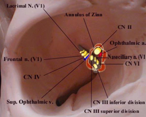
Relationship of nerves and vessels to the annulus of Zinn

Diagram of right orbit that shows the relationship of entering nerves and vessels to the annulus of Zinn.
 The four recti muscles arise from a short funnel-shaped tendinous ring called the annulus of Zinn which encloses the optic foramen and a part of the medial end of the superior orbital fissure. There are 2 tendons.
The Lower Tendon (of Zinn) is attached to the inferior root of the lesser wing of the sphenoid between the optic foramen and the superior orbital fissure. The lower tendon gives origin to part of the medial and lateral recti and all of the inferior rectus. The Upper Tendon (of Lockwood) arises from the body of the sphenoid, and gives origin to part of the medial and lateral recti and all of the superior rectus muscle. The superior and medial recti muscles are much more closely attached to the dural sheath of the optic nerve. This fact may be responsible for the characteristic pain which accompanies extreme eye movements in retro-bulbar neuritis.

Labels: , ,

Monday, December 6, 2010

Corneal Abrasion:What to do and What not to do!

The patient may complain of eye pain or a foreign body sensation after being poked in the eye with a finger or twig. The patient may have abraded the cornea inserting or .................

Labels: ,

Tuesday, November 30, 2010

Glaucoma Surgery 3D Animation

Glaucoma surgery is explained using 3D computer animations

Labels:

Friday, November 26, 2010

Blepharospasm

Blepharospasm is an abnormal tic, spasm, or twitch of the eyelid. It is sometimes referred to as Benign Essential Blepharospasm. Focal dystonia is another phrase for this condition, which involves a involuntary muscle contraction around the eyes. The cause can be fatigue, irritant or caffeine. The symptoms can last for a few days, but commonly disappear without treatment. Severe cases can be chronic.

Besides the tic, symptoms of blepharospasm are eye dryness, excessive blinking, twitching, sensitivity to the sun and bright lights. Blepharospasm occurs spontaneously with no indicators. Patients who experience blepharospasm usually have a history of dry eyes and photo sensitivity. Other syndromes such as Meige’s syndrome have been associated with blepharospasm. Drugs used to treat Parkinson’s disease have also been known to cause blepharospasm. Rarely, blepharospasms can be caused by concussions.

Labels: , ,

Sunday, November 14, 2010

HORDEOLUM

A hordeolum is a common disorder of the eyelid. It is an acute focal infection (usually staphylococcal) involving either the glands of Zeis (external hordeola, or styes) or, less frequently, the meibomian glands (internal hordeola).
Hordeolum (Sty): Infection involving hair follicle of the eyelash.

Internal hordeolum (relatively large, affecting the meibomian glands; may point toward the skin or toward the conjunctive) and external hordeolum (also known as a "sty;" smaller and more superficial; an infection of the glands of Moll or Zeiss; painful; always points toward the skin side of the lid margin).

Pathophysiology
There is usually underlying meibomitis with thickening and stasis of gland secretions with resultant inspissation of the Zeis or meibomian gland orifices. Stasis of the secretions leads to secondary infection, usually by Staphylococcus aureus. Histologically, hordeola represent focal collections of polymorphonuclear leukocytes and necrotic debris (ie, an abscess).


Hordeola should not be confused with chalazia, which represent focal, chronic, lipogranulomatous inflammation of the Zeis or meibomian glands. Chalazia form when underlying meibomitis results in stasis of gland secretions, and the contents of the glands (sebum) are released into the tarsus and adjacent tissues to incite a noninfectious inflammatory reaction. Histologically, chalazia appear as a granulomatous reaction (ie, histiocytes, multinucleated giant cells) surrounding clear spaces that were once occupied by sebum/lipid before they were dissolved by the solvents used for tissue processing, hence the term lipogranuloma.

Both types of hordeola are treated with warm compresses for 10-15 minutes 3-4 times a day; if the condition does not improve within 48 hours, incision and drainage of the pus is indicated. Antibacterial ophthalmic ointment is also helpful.

Labels: ,

Saturday, November 13, 2010

Eye Essentials

This title is directed primarily towards health care professionals outside of the United States. Eye Essentials is a major series which provides authoritative and accessible information for all eye care professionals, whether in training or in practice. Each book is a rapid revision aid for students taking higher professional qualifications and a handy clinical reference guide for practitioners in busy clinics. Highly designed with synoptic text, handy tables, key bullet points, summaries, icons and stunning full colour illustrations, the books have rapidly established themselves as the essential eye clinic pocket books.

Labels: ,

Tuesday, November 9, 2010

Xanthelasma

Xanthelasma are tiny (1-2 mm) yellowish plaques that are slightly raised on the skin surface of the upper or lower eyelids. Xanthelasma is caused by tiny deposits of fat in the skin and is often associated with abnormal blood fat levels (hyperlipidemia).

Xanthelasma typically appears in or near the eyelids. Under the microscope, Xanthelasma can be seen to be composed of lipid-laden foam cells. These cells, termed histiocytes, contain lipid material in their cytoplasm (the nonnuclear zone of the cell). Xanthelasma may be associated with hypercholesterolemia.

Labels: , ,

Saturday, October 23, 2010

Intraocular Lens Implant

Labels:

Sunday, October 17, 2010

Lisch Nodules

 Lisch nodules are melanocytic hamartomas, usually clear yellow to brown, that appear as well defined, dome shaped elevations projecting from the surface of the iris. They may be seen without magnification but a slit lamp examination may be necessary to distinguish them from naevi on the iris which present as flat or minimally elevated, densely pigmented lesions with blurred margins. The nodules are thought not to cause any ophthalmologic complication.

Lisch nodules are the most common clinical finding in adults over 20 years of age with type I neurofibromatosis (NF-1). Unlike cafe au lait spots, multiple nodules are specific for peripheral neurofibromatosis - Von Recklinghausen disease. They may occur in segmental neurofibromatosis which is due to a somatic mutation of the NF-1 gene but are generally absent in central neurofibromatosis. In children, nodules are a more likely finding than neurofibromas. This is an example of an ocular manifestation of a genetic disease. NF-1 has been mapped to a gene defect in chromosome 17.

Labels: ,

Friday, October 8, 2010

Burns of lid margin and superficial cornea

This type of superficial burn injury usually responds to conservative treatment with cycloplegics, debridement as indicated,antibiotic drops and ointment, and adequate pain control.
Scarring is usually minimal as long as Bowman's membrane has not been damaged. Cycloplegia is absolutely essential to control pain from iris reaction to light and to prevent iridocyclitis.

Labels: ,

Sunday, October 3, 2010

Kayser-Fleischer Ring


This is sometimes called as "copper ring" since it appears as a brownish-green copper deposit on the inside surface of the peripheral cornea. It is associated with Wilson disease, a failure to metabolize copper properly.The upper pole is affected more frequently than the lower.

Kayser-Fleischer rings are noted in 90% of patients with Wilson disease and occasionally, in patients with prolonged cholestasis or cryptogenic cirrhosis. The ring is noted most easily in patients with blue eyes. In other patients, slit-lamp examination may be required.
A corresponding gene defect has been mapped to chromosome 13. This is an example of an ocular manifestation of a genetic disease.

Labels: ,