Friday, January 14, 2011

Basosquamous Carcinoma in the Temple


This photo shows  large, crusted tumor on the temple that has clinical features of both squamous cell carcinoma (scaly crust), and basal cell carcinoma (translucency in some areas)........

Read more..........>>

Labels: , ,

Friday, December 31, 2010

Ramsay Hunt Syndrome(clinical)

CAUSES:
Classic Ramsay Hunt syndrome is ascribed to infection of the geniculate ganglion by herpesvirus 3 (varicella-zoster virus [VZV]).


HISTORY:
*Patients usually present with paroxysmal pain deep within the ear. The pain often radiates outward into the pinna of the ear and may be associated with a more constant, diffuse, and dull background pain.

*The onset of pain usually precedes the rash by several hours and even days.

*Classic Ramsay Hunt syndrome can be associated with the following:
-Vesicular rash of the ear or mouth (as many as 83% of cases),The rash might precede the onset of facial paresis/palsy.
-Ipsilateral lower motor neuron facial paresis/palsy (CN VII)
-Vertigo and ipsilateral hearing loss (CN VII)
-Tinnitus,Otalgia,Headaches,DysarthriaGait,ataxia.
-Fever,Cervical adenopathy.

*Facial weakness usually reaches maximum severity by one week after the onset of symptoms.

*Other cranial neuropathies might be present and may involve cranial nerves (CNs) VIII, IX, X, V, and VI.

*Ipsilateral hearing loss has been reported in as many as 50% of cases.

*Blisters of the skin of the ear canal, auricle, or both may become secondarily infected, causing cellulitis.


EXAMINATION:

.The primary physical findings in classic Ramsay Hunt syndrome include peripheral facial nerve paresis with associated rash or herpetic blisters in the distribution of the nervus intermedius.
.The location of the accompanying rash varies from patient to patient, as does the area innervated by the nervus intermedius. It may include the following:
1.Anterior two thirds of the tongue
2.Soft palate
3.External auditory canal
4.Pinna
.The patient may have associated ipsilateral hearing loss and balance problems.
.A thorough physical examination must be performed, including neuro-otologic and audiometric assessment.

Labels: , ,

Tuesday, December 28, 2010

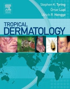
Tropical Dermatology

Stephen K. Tyring, Omar Lupi, Ulrich R. Hengge - Tropical Dermatology
Ch r ,ch .l L g s to .e | ISBN: 0443067902 | 2005-05-06 | PDF | 528 pages | 134 Mb

This is a guide to the mucocutaneous manifestations of tropical diseases. Introductory chapters offer an overview of syndromal tropical dermatology and discuss issues for travelers and those working in the tropics. Protozoa, helminths, viral infections, fungal infections, bacterial infections, and ectoparasitic diseases are then covered, including discussions of epidemiology, diagnosis, differential diagnosis, pathology, laboratory tests, management, and prevention. Non- infectious conditions are also considered in chapters on nutritional diseases, fogo selvagem, pigmentary disorders, and environmental causes of dermatitis.

Labels: ,

Friday, December 24, 2010

Actinic Keratosis of Scalp


This photo shows An actinic keratosis of the scalp. These lesions are often difficult to see.
Description: They are rough, sandpaper-like plaques occurring on sun-exposed surfaces. They are pre-malignant, since a small percentage of them develop into squamous cell carcinoma. The lesion here is darker than the surrounding skin; the lighter area is from the camera flash.

Labels: ,

Saturday, December 18, 2010

Cystic Acne


Cystic Acne is a severe acne with the formation of cysts enclosing a mixture of keratin and sebum in varying proportions. Cystic acne is more likely to leave scars and should be treated by a dermatologist.

FOR MORE CLICK HERE

Labels: ,

Wednesday, December 15, 2010

Alopecia Totalis PIC

Alopecia Totalis (AT): AT is an auto-immune disorder which results in the total loss of hair, but only on the scalp. It is somewhat of an intermediary condition between Alopecia Areata which is patchy scalp hair loss, and Alopecia Universalis which extends to total body hair loss. AT usually appears in two types: One being a fairly sudden and complete loss of all head hair. The other being a slower form which originates as Alopecia Areata (patchy loss) and advances to complete scalp hair loss. In this sense it is sometimes tied to Alopecia Areata (patchy loss), but not all the time.Patients with alopecia areata lose hair on their scalp in smooth round patches typically causing bald spots about an inch (2cm) across.

Most sufferers are children and young adults under the age of 40, though it can affect people of all ages. It can also affect the the nails, giving them a ridged, pitted or brittle appearance. According to statistics, 2% of men and 1% of women in western society suffer from some form of Alopecia Areata. About 2% of those have Totalis or Universalis. This means that about 1 in every 125,000 men and 1 in every 250,000 women have Alopecia Totalis or Universalis.


The main treatment for Alopecia Totalis are therapies which focus on immunomodulation, such as glucocorticoid injections, anthralin, or glucocorticoids taken orally. We have heard reports from some that years of steroid therapy can put the condition into remission. Years of steroid therapy is not always enjoyable however, as there are side effects. It is important to comment that Rogaine (Minoxidil) is not effective for those with Alopecia Totalis. Some treatments which have been considered include Methotrexate, a treatment for autoimmune disorders, and corticosteroids have been proposed as treatments.

Several genes have been studied and quite a bit of research has focused on the human leukocyte antigen. Two studies demonstrated that human leukocyte antigen DQ3 (DQB1*03) was found in more than 80% of patients with Alopecia Areata, which suggests that it can be a marker for general susceptibility to Alopecia Areata. The studies also found that HLA DQ7 (DQB1*0301) and human leukocyte antigen DR4 (DRB1*0401) were found quite a bit more often in patients with Alopecia Totalis (AT) and Alopecia Universalis (AU).

NOTE THAT:
1-alopecia totalis is not a painful disease and does not make people feel sick.
2-It is not contagious, and people who have the disease are usually otherwise
healthy.
3-It has no effect on life expectancy, and will not interfere with your ability to
pursue a normal lifestyle.
4-Returning to your faith can play a major role in understanding what is going on.

Labels: ,

Monday, December 13, 2010

Ephelides (freckles)


Ephelides (also called freckles) are tanned macules seen on the skin. Ephelides are usually multiple in number. Although ephelides are predominantly benign, they may be found in association with systemic disease. Ephelides are associated with fair skin and red or blonde hair. In contrast to solar lentigines, ephelides are not strongly related to age.

Freckle Dermatology The most common pigmented lesion of young light-skinned Caucasians, often of Celtic stock, consisting in light brown 1-10 mm macules which fade in winter, and are accentuated in summer.

Clinically : Ephelides appear during childhood as scattered areas of increased pigmentation, mainly limited to the body regions above the waist.
* These macules are asymptomatic, more numerous on sun-exposed areas, and fade and become smaller in the winter.

On examination, Simple ephelides appear as multiple, small, tanned macules, ranging from 1-5 mm in diameter, with uniform pigmentation. They are most commonly found on sun-exposed areas, such as the nose, the cheeks, the shoulders, and the upper part of the back. The macules may be discrete or confluent.
* Sunburn freckles present similarly to that of simple freckles, but they are darker, have irregular borders, and may be as large as a few centimeters.

Labels: ,

Sunday, December 12, 2010

Appropriate treatment of Rosacea

A 72-year-old man presents to your office complaining of an area of redness associated with the perinasal region. He states that the rash is often worse in the summer, and he has noticed that sunlight exposure makes it worse. Appropriate treatment of this condition consists of
  • A) hydrocortisone cream
  • B) tretinoin gel
  • C) metronidazole cream
  • D) mupirocin ointment
  • E) acyclovir ointment

The answer is: ( C ). (Rosacea)
Rosacea is a common problem encountered by family physicians. The condition is associated with areas of erythema and telangiectasia on the face. It is exacerbated by sunlight, hot or spicy foods, and alcohol. Pronounced rosacea may appear as acneiform papules, pustules, or ruddiness.
Northern Europeans and those of Celtic descent are most commonly affected. Treatment involves oral tetracycline or doxycycline. Topical metronidazole is also effective for milder cases.

Labels: ,

Monday, December 6, 2010

Mucocutaneous Manifestations of Necrolytic Migratory Erythema

Necrolytic migratory erythema is a rare inflammatory dermatosis.which occurs in 70% of patients with the glucagonoma syndrome.
Its clinical features are polymorphic mucocutaneous manifestations, encompassing multiple annular erythematous scaling and crusting patches with hyperpigmentation. These lesions can affect the entire body with a predilection for the perineum, buttocks, groin, abdomen, and limbs — in other words, areas that are subject to increased pressure and friction. The only two reported oral findings are angular cheilitis and atrophic glossitis.
This Figure shows Mucocutaneous Manifestations of Necrolytic Migratory Erythema.
Bullous, erosive oral mucositis with areas having lichenoid features are visible on the tongue and the upper and lower lips (Panel A) and inside the right cheek (Panel B). The patient's entire body, including the back, is covered with purplish, confluent erythematopapulous scaling and crusting lesions with a keratotic surface (Panel C). Histologic analysis of the skin (Panel D, left) shows hyperparakeratosis and spongiosis, accompanied by the presence of necrotic keratinocytes, and vascular proliferation. Histologic analysis of the oral mucosa (Panel D, right) shows a prominent neutrophilic, eosinophilic, and plasmacellular infiltrate (hematoxylin and eosin).

The patient's nutritional status was normal. Routine hematologic tests revealed mild hyperglycemia, hypoalbuminemia, a glucagon level of 73 pg per milliliter (normal range, 25 to 250), and a zinc level of 134 μg per deciliter (normal range, 60 to 250). Tumor markers showed a CA-125 level of 232 U per milliliter (normal range, 0 to 35) and a β2-microglobulin level of 2924 ng per milliliter (normal range, 600 to 2600). Total-body computed tomography revealed the presence of an ovoid mass measuring 40 mm in diameter in the upper lobe of the right lung, with prominent striae hooking the apical and costal homolateral pleura. No evidence of a pancreatic tumor was detected. Needle biopsy of the lung with ultrasonographic guidance revealed a small-cell lung cancer. A diagnosis of nonglucagonoma-associated necrolytic migratory erythema was made. The patient died 2 months later.

Labels: , ,

Saturday, December 4, 2010

Coagulation of Telangiactasia and Excision of Nasal Skin Lesions

Labels:

Friday, November 26, 2010

About head lice infestations

Which of the following statements is true regarding head lice infestations?
  • A) Females are more likely affected.
  • B) Retreatment with pyrethrin is rarely needed.
  • C) Head lice can live off the body up to 1 week.
  • D) Low socioeconomic children are more likely affected.
  • E) Dogs are a common vector for head lice.

The answer is:  ( A ).
Head lice are thought to be the most common type and are developing resistance to commonly used pediculicides. Every year, between 6 million and 12 million people in the United States, primarily children 3 to 10 years of age, are infested with head lice. Girls are at greater risk  because...........

Read more...........>>

Labels: , ,

Thursday, November 25, 2010

Man with characteristic penis  projections

A 41-year-old man complains of soft, raised, flesh-colored growths or projections on his glans penis,prepuce, and penile shaft. Several excisional biopsies are done to look for malignancy.the most likely diagnosis is?
  • a.Genital herpes
  • b.Condyloma acuminatum
  • c.Molluscum contagiosum
  • d.Condylomata lata
  • e.Peyronie’s disease

 The answer is:  ( b ).
The lesions of condyloma acuminatum or venereal warts are soft, flesh-colored (may also be pink or red) growths or .................

Labels: , ,

Sunday, November 21, 2010

Generalized skin hyperpigmentation: causes

"With generalized, none of skin is SPARED ":
  • Sunlight
  • Pregnancy
  • Addison's disease
  • Renal failure
  • Excess iron (haemochromatosis)
  • Drugs (eg busulphan)


Labels: ,

Friday, November 19, 2010

Pachyderma due to scabies

A 65-year-old white man with a history of multiple myeloma presented with thick, leathery, gray skin of the torso and extremities. Panel A shows the left axilla. The patient reported intense pruritus and thickening of his skin during the previous 6 months, despite treatment with oral and topical corticosteroids. Because of chronic immunosuppression due to his underlying malignant condition, a specimen obtained from scrapings of the skin was prepared and examined. It showed scabies mites, eggs, and scybala (fecal pellets) (Panel B), which confirmed a diagnosis of crusted scabies.

The patient was treated with oral ivermectin and topical permethrin, with noticeable improvement within 1 month; subsequently, the patient was lost to follow-up.
Crusted scabies is a rare variant of scabies and occurs most commonly in immunosuppressed patients.
It presents as: erythematous or gray hyperkeratotic patches or plaques that can resemble the hide of a pachyderm (elephant, rhinoceros, or hippopotamus); “pachyderma” refers to thick skin, like that of a pachyderm. Patients with crusted scabies have a very high burden of mites and are extremely infectious. Thus, a prompt diagnosis is needed to avoid transmission to others. No such transmission was documented in this case.

Labels: , , ,

Tuesday, November 16, 2010

Rosacea

A 72-year-old man presents to your office complaining of an area of redness associated with the perinasal region. He states that the rash is often worse in
the summer, and he has noticed that sunlight exposure makes it worse.
Appropriate treatment of this condition consists of :

  • A)hydrocortisone cream
  • B)tretinoin gel
  • C)metronidazole cream
  • D)mupirocin ointment
  • E)acyclovir ointment

The answer is : ( C ).
(Rosacea) Rosacea is a common problem encountered by family physicians. The condition is associated with areas of erythema and telangiectasia on the face. It is exacerbated by sunlight, hot or spicy foods, and alcohol. Pronounced rosacea may appear as acneiform papules, pustules, or ruddiness. Northern Europeans and those of Celtic descent are most commonly affected. Treatment involves oral tetracycline or doxycycline. Topical metronidazole is also effective for milder cases.

Labels: ,

Tuesday, November 9, 2010

Erythema Multiforme Pic


Erythema multiforme is a type of hypersensitivity (allergic) reaction that occurs in response to medications, infections, or illness. Medications associated with erythema multiforme include .................

Labels: ,

Xanthelasma

Xanthelasma are tiny (1-2 mm) yellowish plaques that are slightly raised on the skin surface of the upper or lower eyelids. Xanthelasma is caused by tiny deposits of fat in the skin and is often associated with abnormal blood fat levels (hyperlipidemia).

Xanthelasma typically appears in or near the eyelids. Under the microscope, Xanthelasma can be seen to be composed of lipid-laden foam cells. These cells, termed histiocytes, contain lipid material in their cytoplasm (the nonnuclear zone of the cell). Xanthelasma may be associated with hypercholesterolemia.

Labels: , ,

Monday, November 8, 2010

Ephelides (freckles)


Ephelides (also called freckles) are tanned macules seen on the skin. Ephelides are usually multiple in number. Although ephelides are predominantly benign, they may be found in association with systemic disease. Ephelides are associated with fair skin and red or blonde hair. In contrast to solar lentigines, ephelides are not strongly related to age.

Freckle Dermatology The most common pigmented lesion of young light-skinned Caucasians, often of Celtic stock, consisting in light brown 1-10 mm macules which fade in winter, and are accentuated in summer.

Clinically : Ephelides appear during childhood as scattered areas of increased pigmentation, mainly limited to the body regions above the waist.
* These macules are asymptomatic, more numerous on sun-exposed areas, and fade and become smaller in the winter.

On examination, Simple ephelides appear as multiple, small, tanned macules, ranging from 1-5 mm in diameter, with uniform pigmentation. They are most commonly found on sun-exposed areas, such as the nose, the cheeks, the shoulders, and the upper part of the back. The macules may be discrete or confluent.
* Sunburn freckles present similarly to that of simple freckles, but they are darker, have irregular borders, and may be as large as a few centimeters.

Labels: ,

Peutz-Jeghers syndrome

A 10 years old girl presents with multiple pigmented macules on the vermilion border of her lower lip.The dark brown lesions are 2–5 mm in size and are arranged in a cluster.The patient’s older brother has similar lesions. The patient complains of recurrent bouts of abdominal pain. the most likely diagnosis is?..............

Read more..............>>

Labels: , ,

Sunday, November 7, 2010

An Atlas of Psoriasis, Second Edition (Encyclopedia of Visual Medicine Se


Completely revised, this second edition is a full update of the internationally acclaimed first edition of An Atlas of Psoriasis, written by Lionel Fry, one of the world’s most distinguished dermatologists. Copiously illustrated in color, including new high quality visual imagery only recently available, the atlas is designed to replace the old edition on the shelves of dermatologists and dermatopathologists worldwide. Like the previous edition, the book provides an authoritative review of psoriasis, including clinical features, sites and clinical patterns, differential diagnosis, psoriatic arthropathy, and treatment.

For Download :

Labels: ,