Thursday, December 30, 2010

Case of gross hematuria following a game of soccer

A 37 year old male complains of gross hematuria following a game of soccer with his friends at work. He could not recall any particularly severe trauma. On examination both kidneys were enlarged and easy palpable. His blood pressure was raised. A likely diagnosis is :

a) polycystic disease of the kidneys
b) Berger's nephritis
c) systemic lupus erythematosus
d) renal vein thrombosis
e) Goodpasture's syndrome

The correct answer is...................... A

Explanation
In polycystic disease of the kidneys the symptoms or signs first occur between the third and fourth decades of life. These include microscopic and gross hematuria, flank pain and hypertension. The polycystic condition is not confined to the kidneys. Hepatic cysts, usually identified incidentally by sonography, help in making the diagnosis of DPK. These cysts are more likely to be found in adults than in children. Approximately 10% to 40% of patients have berry aneurysms, and approximately 9% of these patients die because of subarachnoid hemorrhages

Labels: ,

Wednesday, December 29, 2010

Benign Prostatic Hyperplasia

Many men with benign prostatic hyperplasia experience urinary problems related to the condition. As the prostate enlarges, the gland places increasing pressure on the urethra, often resulting in difficulty beginning or ending urination, an inability to completely empty the bladder, decreased urine flow, and frequent urination. In the most severe cases, complete blockage of the urethra occurs, which may lead to kidney damage.
From microscopyu.com

Benign Prostatic Hyperplasia at 20x Magnification :
Part of the male reproductive system, the prostate gland produces and stores seminal fluids, releasing them into the urethra when semen emission occurs. The gland is located directly below the bladder and surrounds the upper part of the urethra. During adolescence the gland usually matures and reaches a size comparable to that of a walnut. The dimensions of the gland generally remain unchanged for several decades, but in most older men, the prostate begins to enlarge as the size of its cells increases, a process commonly referred to as benign prostatic hyperplasia (BPH) or hypertrophy. According to recent estimates, more than 50 percent of men between the ages of 50 and 60 experience benign prostatic hyperplasia, and over 90 percent of those 70 to 90 years old have developed the condition. Researchers do not yet completely understand the cause of this physiological change, but it is widely thought that elevated levels of the female sex hormone estradiol and increased manufacture of dihydrotestosterone, a derivative of the male sex hormone testosterone, contribute to the condition.

Benign Prostatic Hyperplasia at 4x Magnification :
Men with only mild symptoms of benign prostatic hyperplasia may elect not to undergo any treatment or to simply take a wait-and-see attitude, visiting the doctor regularly for monitoring until signs suggest a more active approach is needed. For those who seek treatment, a number of options are available. For example, drugs such as alpha blockers and finasteride may be used alone or in conjunction with one another to relax prostatic smooth muscle and decrease the size of the prostate gland. Individuals that are not responsive to the typical medications, however, may require a more invasive form of treatment, such as balloon dilation of the urethra or any of several different surgical techniques, including transurethral incision of the prostate (TUIP), transurethral resection of the prostate (TURP), or open prostatectomy. The various treatments for benign prostatic hyperplasia are associated with a number of risks and side effects, which can include serious conditions like incontinence and impotence.

Labels: ,

Monday, December 27, 2010

Transposition of Femal Ureter

Labels:

Tuesday, December 21, 2010

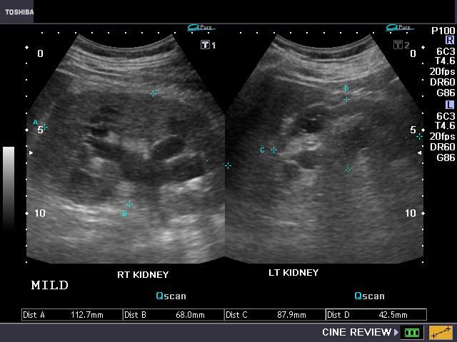
Grades of hydronephrosis (on Ultrasound imaging):

A) Mild hydronephrosis:
This images shows mild dilatation of the pelvis as well as the calyces of the right kidney suggesting mild hydronephrosis. The left kidney also appears to ............


Labels: ,

Friday, December 3, 2010

Minimally Invasive Surgery Corrects Urinary Incontinence

A DMC patient with urinary stress incontinence regains control with less-invasive surgery at DMC Sinai-Grace, performed by DMC Ob/Gyn specialist Dr. Korial Atty. ~ Detroit Medical Center.

Labels: ,

Saturday, October 2, 2010

A Case of left Flank Pain

A 48-year-old woman presented to the emergency department with three days of fever, along with rigors, left flank pain and oliguria. She had diabetic nephropathy with a baseline serum creatinine level of 247.5 (normal < 133) µmol/L and an estimated glomerular filtration rate of 19 (normal 91–130) mL/min/1.73m2. The patient’s temperature was 38.8°C, blood pressure was 139/73 mm Hg and she had tenderness at the left costovertebral angle.
Laboratory studies
revealed a hemoglobin concentration of 90 (normal 120–160) g/L, leukocyte count 31.48 (normal 4.3–10.8) x 109/L, platelet count 296 (normal 130–400) x 109/L and hemoglobin A1C of 8.5%. Serum creatinine was 530.4 (normal < 133) µmol/L and urinalysis showed 5–10 white blood cells per high-power field. A plain radiograph showed a rosette-like arrangement of gas in the collecting system of the patient’s left kidney and upper ureter . An unenhanced computed tomography (CT) scan of the abdomen showed air in the dilated renal calyces, pelvis and upper ureter . No evidence of urinary tract obstruction was found. The patient was diagnosed with emphysematous pyelitis. Peripheral blood and urine cultures grew Escherichia coli, sensitive to ceftriaxone. (A) Plain radiograph of a 48-year-old woman showing rosette-like arrangement of gas in the collecting system of her left kidney (white arrows) and upper ureter (black arrow). (B) Coronal view of an abdominal computed tomography scan showing air in the dilated renal calyces, pelvis and upper ureter (white arrows). A tiny renal stone (black arrow) is present.

Treatment with nephrostomy and intravenous ceftriaxone 2 g daily led to clinical and radiological improvement. On follow-up at three weeks, the patient’s creatinine level was 265.2 µmol/L and estimated glomerular filtration rate 18 mL/min/1.73m2.

What we learned??
  1. Emphysematous pyelonephritis occurs more commonly in middle-aged women with poorly controlled diabetes or urinary tract obstruction. It should be considered in patients with pyelonephritis who develop thrombocytopenia, acute renal failure, reduced level of consciousness or shock. These factors also contribute to increased mortality.
  2. Escherichia coli is the most common organism found in emphysematous pyelonephritis, followed by Klebsiella pneumoniae.
  3. Ultrasonography with plain radiography is recommended in all patients with diabetes who have acute pyelonephritis. Imaging is not generally recommended in patients who are otherwise well who respond to therapy within 72 hours.
  4. Computed tomography is the most sensitive method to demonstrate gas within and adjacent to the kidney.
  5. The initial management of emphysematous pyelonephritis includes resuscitation, antibiotic treatment targeting Gram-negative bacteria and control of diabetes if present.
  6. Most patients require nephrostomy. Emphysematous pyelitis has an excellent prognosis with prompt diagnosis and treatment.

Labels: ,

Tuesday, September 28, 2010

Emergencies in Urology

Emergencies in Urology
Publisher: Springer | ISBN: 3540486038 | edition 2007 | PDF | 679 pages | 41,5 mb


Emergencies in Urology is a comprehensive textbook covering one of the few remaining white spots on the map of urological literature. To date only a small number of publications have been dedicated to the topic of urgent and emergent problems in urology – important as these are in our daily clinical life.Therefore the editors, both of them internationally recognized urological experts, have taken the effort to present an in-depth study into virtually every possible urgent urological situation with which a urologist may be confronted today. Consequently, the book includes chapters on topics such as urological trauma, urosepsis, urinary obstruction, oncological emergencies, intra- and postoperative complications, acute problems in children, and many more.To obtain the best possible expertise in such a wide field, renowned expert authors have contributed their experience to this book. They did so not only by writing the regular book chapters but also by delivering short stories about urgent situations they encountered in their own professional life. These vignettes are one of the rare opportunities where experience can be relayed without restriction from one urological generation to the next.Much work has gone into the illustrations for the book. Foremost in this respect is the art of Stephan Spitzer, one of the leading medical illustrators of today.The result is a comprehensive, well-organized text, in which state-of-the-art know-how, didactic algorithms, personal experience and detailed illustrations are combined into a unique guide of how to manage urological emergencies.

For Free Download:

Labels: , ,

Tuesday, September 7, 2010

Urinary catheters :Do's and don'ts


It is a form of drain which Can be inserted transurethrally or suprapubically
*Commonly used to:
1- Alleviate or prevent urinary retention
2- Monitor urine output

* Catheters vary by:
o The material from which they are made (latex, plastic, silastic, teflon-coated)
o The length of the catheter (38 cm 'male' or '22 cm 'female')
o The diameter of the catheter ................

Labels: , , ,

Urinary tract infection

A urinary tract infection (UTI) is a bacterial infection that affects any part of the urinary tract. The main causative agent is Escherichia coli. Although urine contains a variety of fluids, salts, and waste products, it usually does not have bacteria in it. When bacteria get into the bladder or kidney and multiply in the urine, they cause a UTI. The most common type of UTI is a bladder infection which is also often called cystitis. Another kind of UTI is a kidney infection, known as pyelonephritis, and is much more serious. Although they cause discomfort, urinary tract infections can usually be quickly and easily treated with a short course of antibiotics.Symptoms include frequent feeling and/or need to urinate, pain during urination, and cloudy urine.

/>

Labels:

Thursday, September 2, 2010

Atlas of Urologic Surgery


B. Saunders. Univ. of Calfornia School of Medicine, San Francisco. Atlas of surgical technique for urologists. Extensive halftone line drawings with commentary.

PART 1

PART 2

Labels: ,

Thursday, August 26, 2010

Hutch diverticula of Bladder

A 73-year-old man presented to the emergency department after having intermittent fevers for 2 weeks. He had a history of recurrent urinary tract infections, parkinsonism, and a compression fracture at the L2 vertebra that was the result of a fall 2 years before presentation.
In addition, he had paraparesis and a neurogenic bladder, also subsequent to the fall. The results of physical and laboratory evaluation were notable for the identification of Pseudomonas aeruginosa in a blood culture and for a urinalysis showing more than 100 white cells per high-power field. He was treated with empirical antibiotics.

Intravenous urography showed no obstructive uropathy, but symmetric diverticula could be seen near both ureteral orifices (arrows). These lesions, known as Hutch diverticula, are usually congenital rather than occurring as a result of a neurogenic bladder or an infection or obstruction. They represented a new finding in this patient. Hutch diverticula are more commonly seen in men and boys and are usually unilateral and asymptomatic. After treatment with antibiotics, the patient's fever and pyuria subsided.
He declined any further evaluation or intervention. During the year after diagnosis, two more urinary tract infections developed.

Labels: ,

Monday, July 26, 2010

LITHOTRISY SIDE EFFECTS

Labels:

Monday, July 19, 2010

PROSTATE Cancer Explanation [3D]

Labels:

Saturday, July 17, 2010

LITHOTRISY SIDE EFFECTS

Labels: ,

WHAT IS LITHOTRIPSY

Labels: ,

Tuesday, July 6, 2010

The Largest Kidney Stone

Thursday, June 3, 2010

3- D ultrasound image WOW !!

These 3 dimensional ultrasound images show various pathologies of the urinary bladder, providing a whole new "dimension" to sonographic imaging.

3- D ultrasound image of ureterocele:
The Rt. ureterocele is seen as a small sac bulging in from the posterior wall of the bladder.


3-D ultrasound image of bladder diverticulum:
A small right sided vesical diverticulum. (UB= urinary bladder)


Vesical calculus:
This 3-D ultrasound image of a vesical calculus shows that 3-D imaging must be used in conjunction with 2-D B-mode imaging to identify pathology. This bladder calculus is difficult to distinguish from (see topmost image) ureterocele purely on 3-D ultrasound. Perhaps a cross sectional image would show the internal structure better. (All images courtesy of Ravi Kadasne, MD, UAE).

Labels: ,

Monday, May 31, 2010

Foley's catheter insertion in male and female

Saturday, May 29, 2010

Prostatectomy :Fantastic 3D Animation

Labels:

Friday, May 21, 2010

Calcifications in a Continent Urinary Diversion


A 37-year-old man with a history of spina bifida presented with an audible knocking sound coming from his abdomen during ambulation. Fifteen years earlier he had undergone a cystectomy with a continent cutaneous urinary diversion (Indiana pouch) for a neurogenic bladder. He had been poorly compliant with the catheterization and irrigation regimen involving the pouch.
Physical examination revealed a well-healed, functional stoma and a palpable mass in the right side of the abdomen. Urinalysis revealed a urinary pH of 9.0, with numerous white and red cells, and urine culture grew Proteus mirabilis.

Plain abdominal radiography
revealed two large multilaminar calcifications in the right side of the abdomen (Panel A). Computed tomography of the abdomen and pelvis confirmed the presence of two large calculi within the Indiana pouch (Panel B). The patient underwent cystolithotomy without incident. At a follow-up visit 1 year after the procedure, he was found to have been compliant with his catheterization and irrigation regimen and remained free of stones.

Continent urinary diversions may be associated with hypercalciuria, hyperphosphaturia, hyperoxaluria, and hypocitraturia, all of which may predispose patients to pouch urolithiasis. Infections with organisms that break down urea can result in an alkaline environment, which can in turn give rise to struvite (magnesium ammonium phosphate) stones. Establishment of a formal catheterization and irrigation protocol for continent urinary diversions may reduce the risk of urolithiasis.

Labels: ,